WHAT IS PHOTOACOUSTIC IMAGING?
Photoacoustic imaging is a non-invasive imaging technique that combines laser-induced ultrasound waves with traditional optical imaging to visualize structures and functional properties of biological tissues.
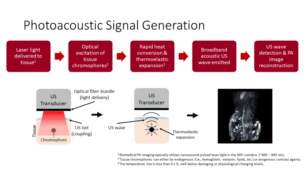
HOW DOES PHOTOACOUSTIC IMAGING WORK?
Photoacoustic imaging (PAI) is a hybrid imaging modality that combines optical imaging and ultrasound imaging (light+sound). ??? It works by using light to excite molecules in tissue, which causes them to heat up and produce sound waves.
These sound waves are then detected by ultrasound transducers and used to create an image of the tissue.
PAI is a relatively new imaging technology, but it has already shown great potential in a variety of applications, including:
• Cancer detection and diagnosis: PAI can be used to detect and characterize tumors, including their size, shape, and location. It can also be used to assess the response of tumors to treatment,
• Cardiology: PAI can be used to image the heart, including the chambers, valves, and vessels. It can also be used to assess the function of the heart, such as its pumping efficiency.
• Dermatology: PAI can be used to image the skin, including the epidermis, dermis, and subcutaneous tissue. It can also be used to assess the severity of skin conditions, such as psoriasis and melanoma.
• Other applications: PAI is also being investigated for use in a variety of other applications, such as dentistry, gastroenterology, and neuroscience.
PAI is a promising new imaging technology with a wide range of potential applications. It is still under development, but it has already shown great potential to improve the diagnosis and treatment of a variety of diseases.
Here are some of the advantages of photoacoustic imaging:
• High contrast: PAI can provide high-contrast images of tissue, even in the presence of blood. This is because the optical absorption of tissue varies with its composition, such as the concentration of blood, oxygen, and other molecules.
• High spatial resolution: PAI can provide high spatial resolution images of tissue, up to a few micrometers. This is comparable to the resolution of MRI and CT scans.
• Non-invasive: PAI is a non-invasive imaging technique, which means that it does not require the use of needles or incisions. This makes it a safe and convenient option for patients.
• Real-time imaging: PAI can provide real-time images of tissue, which can be used to track the changes in tissue as a result of a medical procedure or treatment.
Here are some of the limitations of photoacoustic imaging:
• Depth of penetration: The depth of penetration of PAI is limited by the attenuation of light in tissue. This means that PAI is not well-suited for imaging deep tissues, such as the brain or liver.
• Cost: PAI systems are still relatively expensive, which limits their availability.
• Expertise: PAI is a relatively new technology, and it requires specialized expertise to operate and interpret the images.
As the technology continues to develop, it is likely to become more affordable and accessible, and its use will likely expand to a wider range of clinical applications.