whole body small animal 3D imaging platform
Description
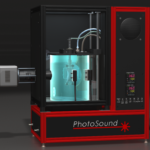
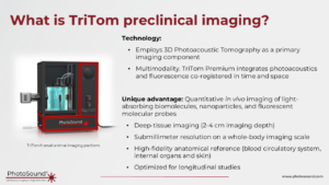
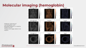
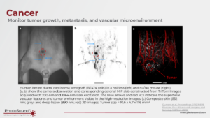
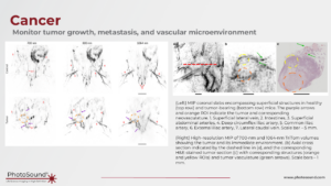
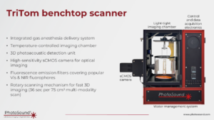
The TriTom imaging platform utilizes photoacoustic and fluorescence tomographies (PAFT) to enable high-resolution (up to 150 µm) non-invasive in vivo whole-body imaging in small animal models. With the ability to use multiple excitation wavelengths per scan, the multi-modality system can simultaneously acquire photoacoustic and fluorescence data in large volumes (30-60 cm3) allowing for spectroscopic molecular analysis within the region of interest. In addition to 3D molecular maps, the TriTom enables spatially-resolved assessment of physiologic parameters in vivo, such as volumetric blood content and oxygenation without the need for contrast agents. The TriTom also provides quantitative imaging for a wide range of fluorophores and other types of molecular probes excited between 460 nm and 1320 nm. With such versatile imaging capabilities, the state-of-the-art TriTom system is ideally suited for a wide range of preclinical applications including cancer, toxicology, developmental biology, tissue engineering and regeneration, neuroscience, cardiovascular imaging, as well as in the development of drugs, therapies, and optical and fluorescent imaging probes.
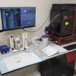
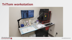
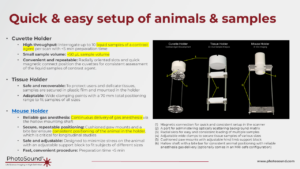
The TriTom is specially designed for imaging small animal models with a convenient animal handling set-up for fast, reproducible positioning of the subject and integrated gas anesthesia to quickly move from animal preparation to imaging. The rapid image acquisition time (~36 seconds) allows for fast imaging of individual subjects or capturing dynamic processes such as biodistribution, lymphatic drainage, or vasodilation. Additionally, the temperature control unit maintains the imaging environment within ± 0.1°C, ensuring the animal’s welfare throughout the imaging session.
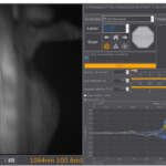
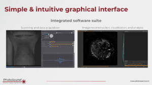
A user-friendly image acquisition software enables new users to start collecting data quickly while maintaining the flexibility for advanced users to optimize imaging parameters for their specific research applications. The image reconstruction software is also designed for rapid reconstruction and visualization of large volumes with an open data format that allows for flexibility in image reconstruction and data management. With such versatile imaging capabilities, the state-of-the-art TriTom system is ideally suited for a wide range of preclinical applications.
Features
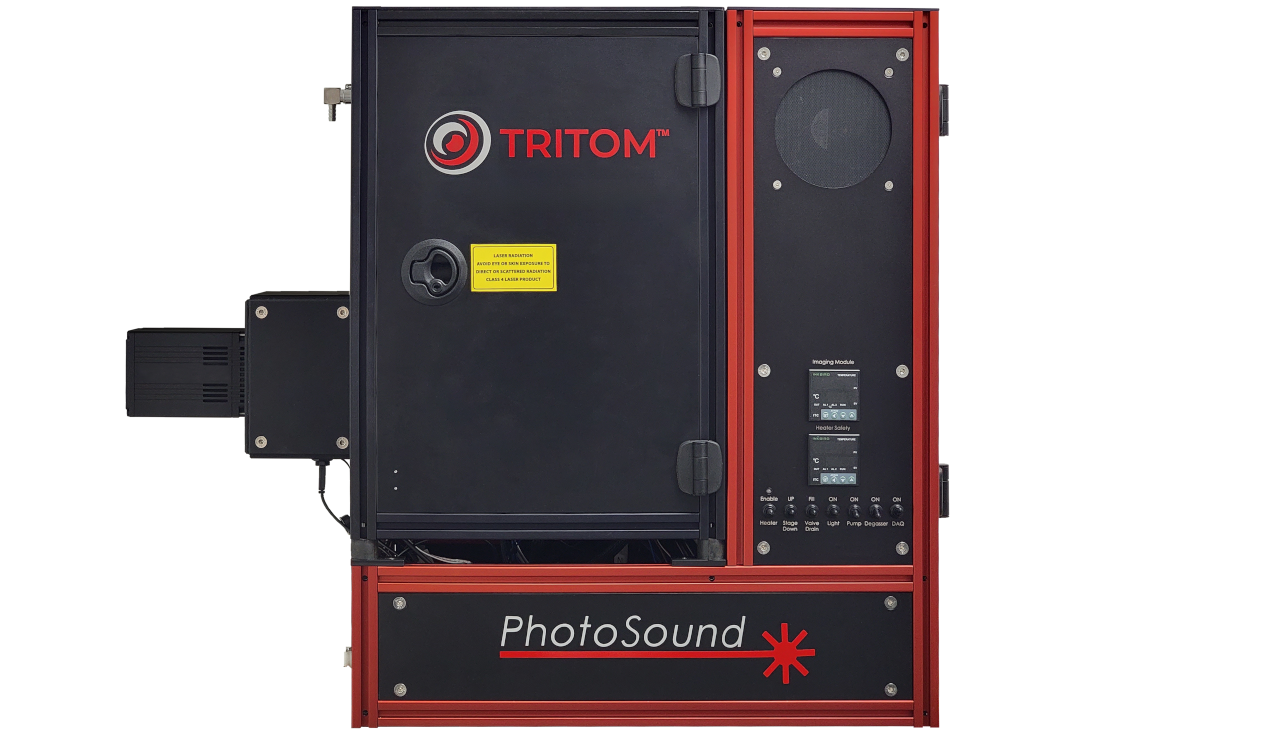
- Combined 3D photoacoustic and fluorescence tomography in a compact benchtop design
- Fast imaging scans (<36 s) of large volumes (30-60 cm3) with superior molecular sensitivity
- Accurate 3D registration of anatomical, functional, and molecular volumes with a high spatial resolution (up to 150 µm)
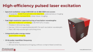
- Tunable optical excitation wavelength across the entire spectral range, enabling multiple wavelengths in a single scan
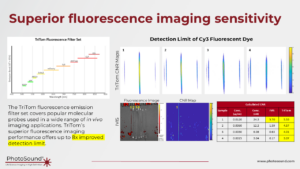
- High-sensitivity sCMOS fluorescence camera with standard emission filters covering popular fluoroprobes
- Integrated gas anesthesia line and adjustable mouse holder for convenient operation and repeatable in vivo longitudinal studies
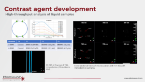
- Single-scan assessment of multiple micro samples (50 µL or less) to accelerate contrast agent development without wasting expensive samples
- User-friendly integrated software suite designed to minimize experiment time and reconstruct large-scale volumes in seconds
- Open data format allowing for image reconstruction and data management with third-party software