Cancer Detection and Monitoring with the TriTomTM
Photoacoustic imaging has become a popular tool in preclinical cancer research as a noninvasive technique for monitoring tumor growth and therapeutic response. Specifically, photoacoustic imaging can provide high-sensitivity images of both superficial and deep vasculature and quantitative assessment of blood oxygen saturation without exogenous contrast. The TriTom small animal imaging platform provides high-resolution photoacoustic tomography (PAT) images, enabling whole-body in vivo anatomical, functional, and molecular analysis for longitudinal cancer studies.
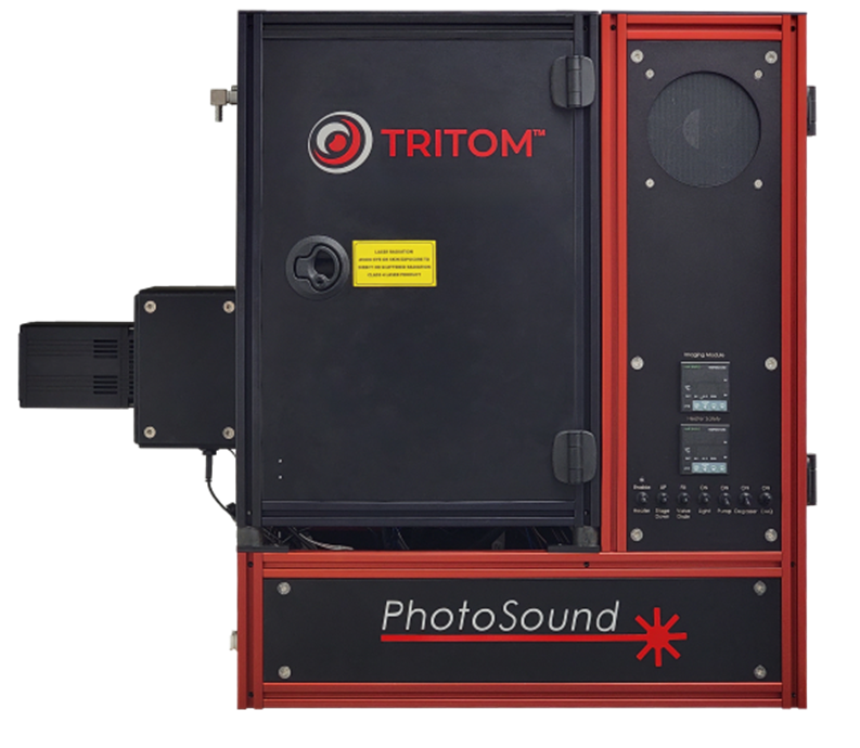
Imaging System TriTomTM
Excitation Wavelengths 460 – 1300nm
Spatial Resolution Up to 160 µm (PA)
Up to 70 µm (FL)
Acquisition Time 36 s per scan
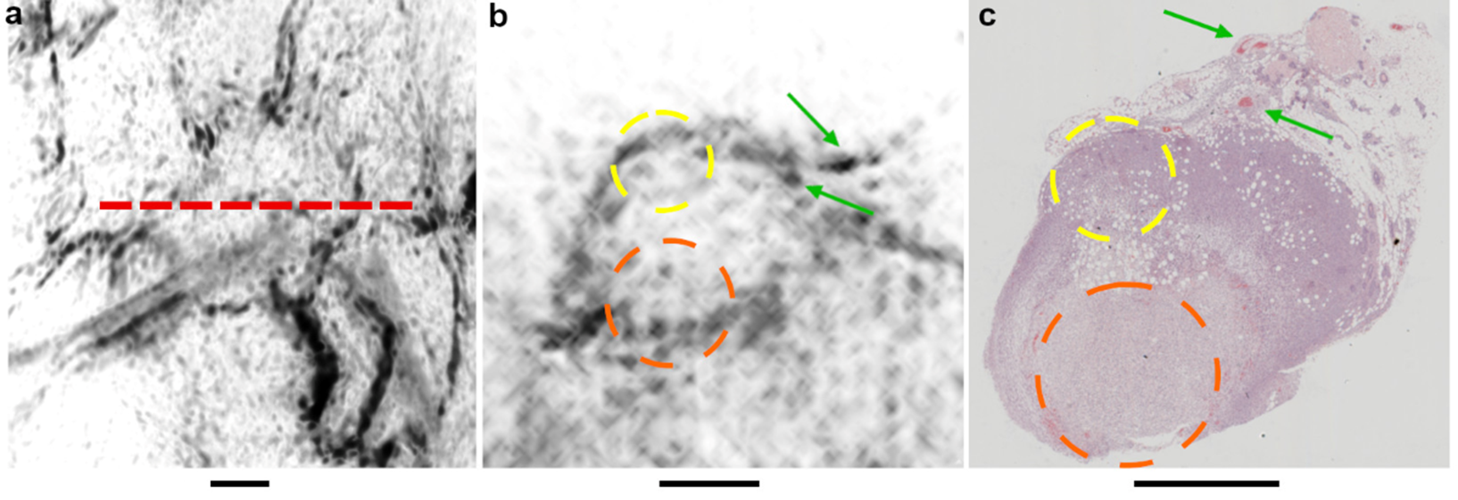
Monitoring Microvascular Development
The microvascular network of a tumor not only regulates the supply of nutrients that contribute to growth and metastasis but influences the response to anticancer therapies. Preclinical studies of tumor growth and neovasculature development are, therefore, critical to fundamental cancer research and therapeutic development. The TriTom is a 3D imaging platform that can resolve the complex and size-varying blood vessels supplying the tumor and quantify the local density of microvessels. Further, the TriTom’s superior spatial resolution in all three anatomical planes allows for visualization and monitoring of the microvascular network from any angle. This unique advantage enables longitudinal monitoring of tumor vasculature development and evaluation of therapies targeting cancer blood supply.
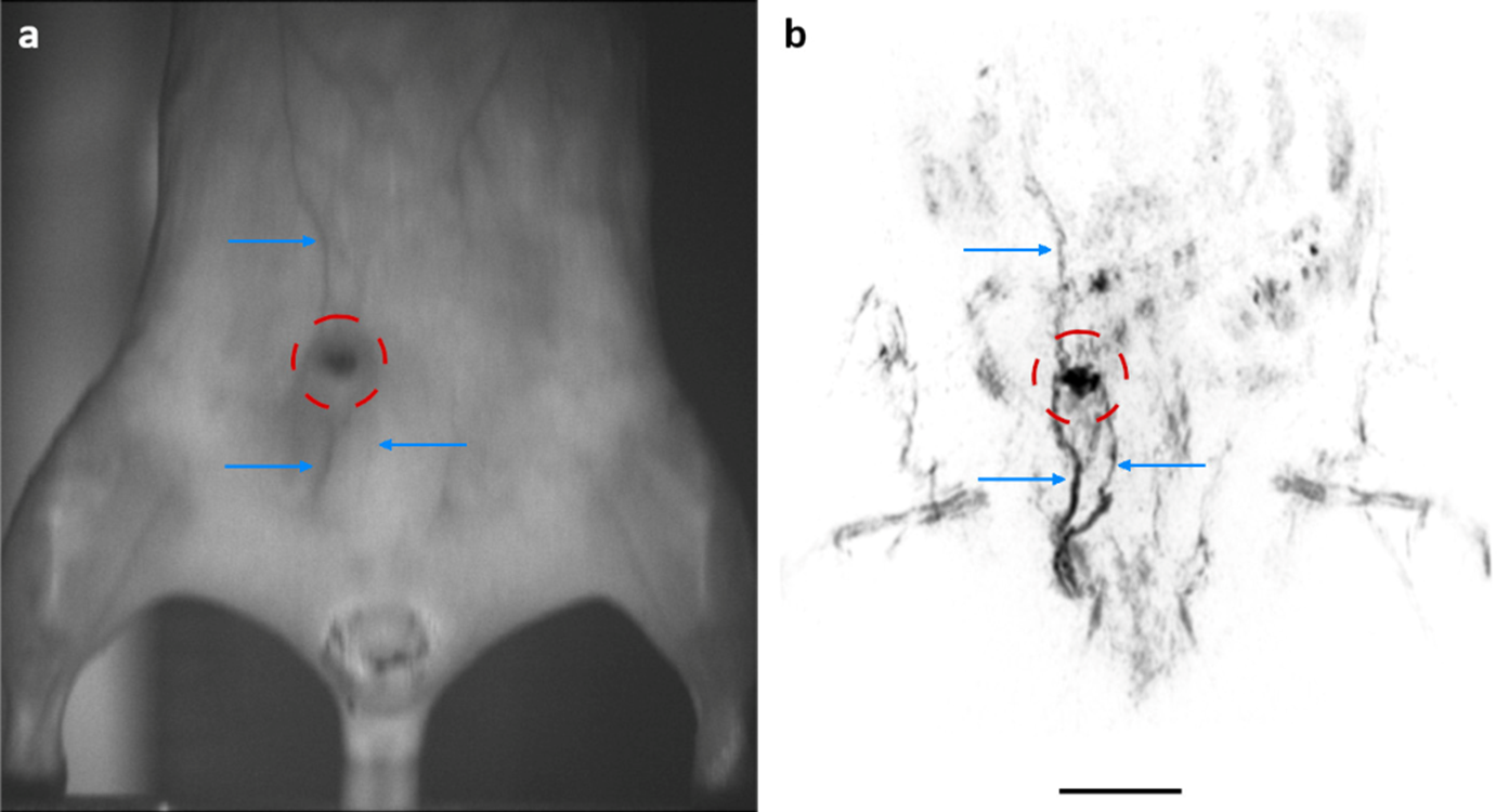
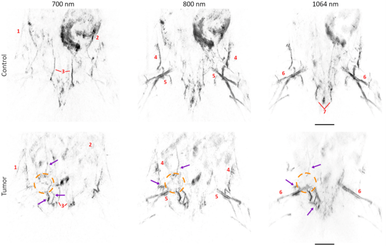
Tumor Growth Monitoring
Accurate noninvasive measurements of tumor size and morphology are crucial to evaluating the effects of novel therapies. However, current modalities provide an incomplete picture of the tumor environment due to poor spatial resolution and limited imaging depth. The TriTom is a high-resolution 3D imaging technology with superior molecular sensitivity in deep tissue. As a result, the TriTom images provide a detailed view of the tumor morphology and enable accurate longitudinal assessment of tumor growth. These key features make the TriTom a powerful tool for monitoring tumor growth, metastasis, and therapeutic response in preclinical cancer research.