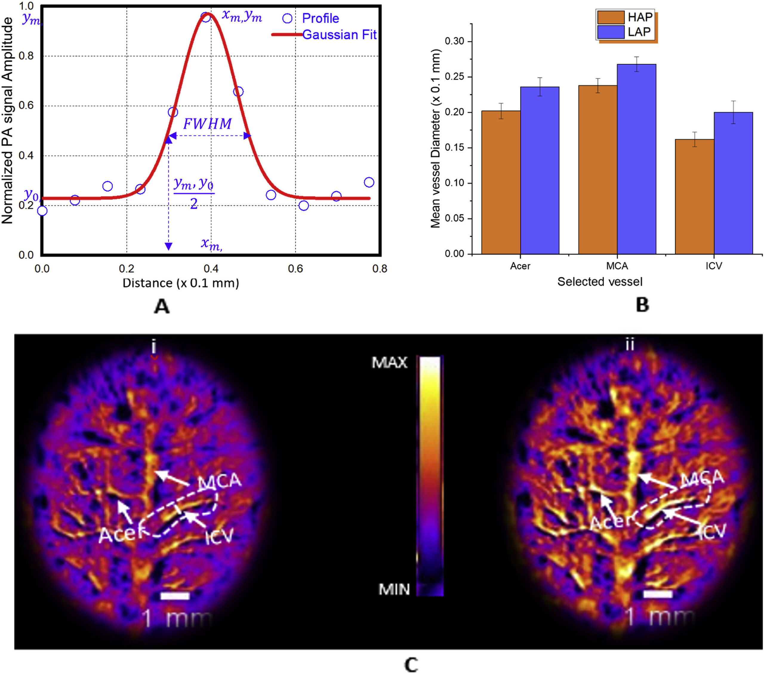
A) Details of a normalized Gaussian fit for one of the vessels of interest from which full width at half maximum (FWHM) is estimated (ICV – inferior cerebral vein; MCA – middle cerebral artery; Acer – anterior cerebral artery). B) Results of statistical analysis and comparison between selected vessels for five HAP mice and five LAP mice (3 × 5 vessels for each animal model). The error bars indicate the standard deviation in the calculation. With a p value of p < 0.002 (F = 40.38), the vessels’ diameters for HAP mice are significantly smaller than for LAP mice. C(i) and C(ii), typical PAT images of HAP and LAP mouse cerebral cortex, respectively, at 800 nm wavelength.