Author(s): Yaocai Huang, Ibrahim Oraiqat, Dale Litzenberg, Madhumithra Subramanian Karthikes, Christopher Tichacek, Glebys Gonzalez, Zhanpeng Xu, Sarah Dykstra, Borui Li, Scott Hadley, Eduardo G. Moros, Man Zhang, Paul L. Carson, Kyle C. Cuneo, Xueding Wang, Issam El Naqa, Wei Zhang
ABSTRACT
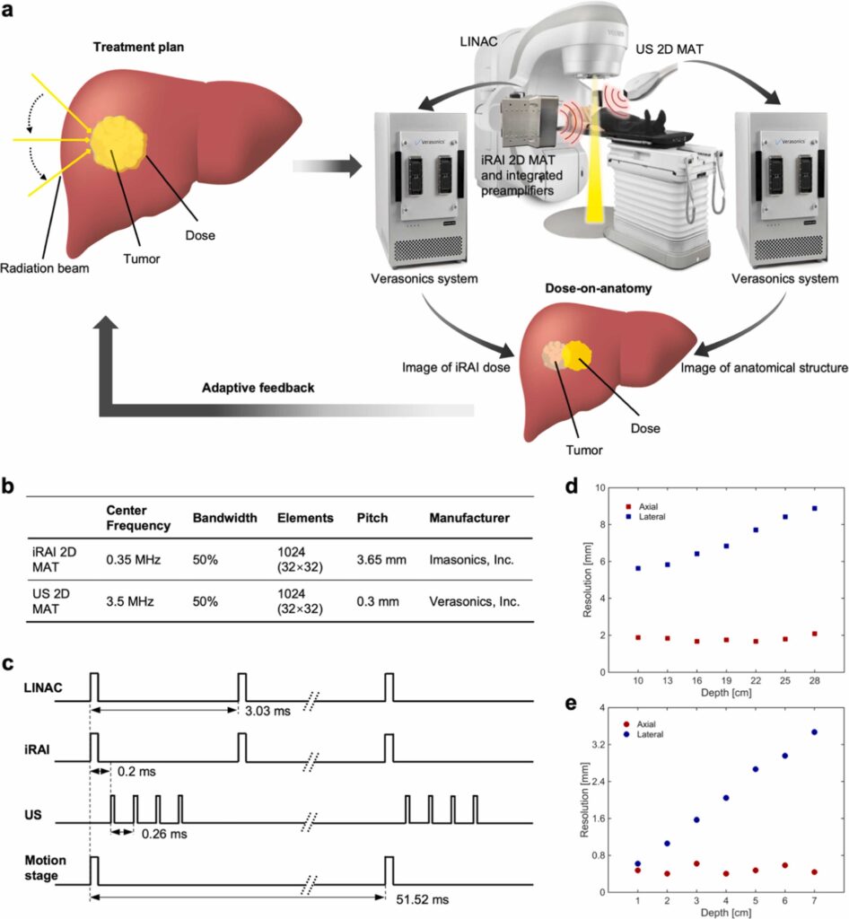
The aim of this study is to visualize the radiation dose on anatomical structures during radiation therapy (RT) by mapping radiation dose deposition and tracking anatomical structures simultaneously. A dual-modality volumetric imaging system, which combines ionizing radiation acoustic imaging (iRAI) and ultrasound (US) imaging, was developed to provide dose deposition and anatomical information in real-time during RT. The performance of the proposed system was first evaluated via experiments on tissue-mimicking phantoms driven by a custom motion stage. By using US imaging to correct the position of anatomical structures, the dose mapping accuracy of the system increased by up to 0.51 in structural similarity index measure (SSIM) and 74.60 % in Gamma passing rate (GPR) compared to standalone iRAI. A subsequent study on a rabbit model in vivo further confirmed the capability of the system in mapping of the radiation dose deposition in the target tissue as well as its change caused by the motion mainly due to the animal breath. These findings demonstrate that this first-of-its-kind dual-modality volumetric imaging system can provide volumetric dose-on-anatomy information during RT. After further validation in clinic, this technique holds potential for enhancing RT outcomes by ensuring accurate alignment between the planned radiation beams, the target, and surrounding organs at risk.