Recent Posts

INDOCYANINE GREEN DYE BASED BIMODAL CONTRAST AGENT TESTED BY PHOTOACOUSTIC/FLUORESCENCE TOMOGRAPHY SETUP

Authors: MAKSIM D. MOKROUSOV, WEYLAN THOMPSON, SERGEY A. ERMILOV, TATIANA ABAKUMOVA, MARINA V. NOVOSELOVA, OLGA A. INOZEMTSEVA, TIMOFEI S. ZATSEPIN, VLADIMIR P. ZHAROV, EKATERINA I. GALANZHA, DMITRY A. GORIN 


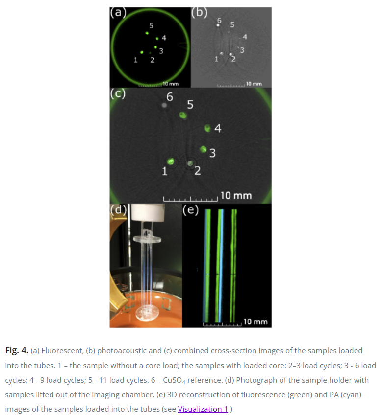
Visualization 1. 3D image of the samples reconstructed from the obtained photoacoustic and fluorescent measurement data
ABSTRACT

Multimodal imaging systems are in high demand for preclinical research, experimental medicine, and clinical practice. Combinations of photoacoustic technology with other modalities including fluorescence, ultrasound, MRI, OCT have been already applied in feasibility studies. Nevertheless, only the combination of photoacoustics with ultrasound in a single setup is commercially available now. A combination of photoacoustics and fluorescence is another compelling approach because those two modalities naturally complement each other. Here, we presented a bimodal contrast agent based on the indocyanine green dye (ICG) as a single signalling compound embedded in the biocompatible and biodegradable polymer shell. We demonstrate its remarkable characteristics by imaging using a commercial photoacoustic/fluorescence tomography system (TriTom, PhotoSound Technologies). It was shown that photoacoustic signal of the particles depends on the amount of dye loaded into the shell, while fluorescence signal depends on the total amount of dye per particle. For the first time to our knowledge, a commercial bimodal photoacoustic/fluorescence setup was used for characterization of ICG doped polymer particles. Additionally, we conducted cell toxicity studies for these particles as well as studied biodistribution over time in vivo and ex vivo using fluorescent imaging. The obtained results suggest a potential for the application of biocompatible and biodegradable bimodal contrast agents as well as the integrated photoacoustic/fluorescence imaging system for preclinical and clinical studies.

Read the full publication →

Citation

M. D. Mokrousov, W. Thompson, S. A. Ermilov, T. Abakumova, M. V. Novoselova, O. A. Inozemtseva, T. S. Zatsepin, V. P. Zharov, E. I. Galanzha, and D. A. Gorin, “Indocyanine green dye based bimodal contrast agent tested by photoacoustic/fluorescence tomography setup,” Biomed. Opt. Express, vol. 12, no. 6, p. 3181, 2021, doi: 10.1364/boe.419461.
GOLD NANOPARTICLES CONJUGATED WITH DNA APTAMER FOR PHOTOACOUSTIC DETECTION OF HUMAN MATRIX METALLOPROTEINASE-9

Authors: Jinhwan Kim, Anthony M Yu, Kelsey P. Kubelick, Stanislav Y. Emelianov


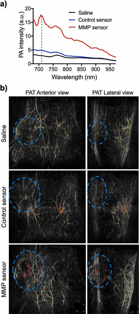
Fig. 5. Spectroscopic analysis and PAT of the MMP sensor in an in vivo mouse model of breast cancer. (a) PA intensity from 680 nm to 970 nm of the MMP sensor in the tumor region and assorted controls – saline only and control sensor. Black dashed line marks 710 nm. (b) Overlaid photoacoustic tomography of saline, control sensor, and MMP sensor-injected mice. The volumes of skin and surface vasculature (532 nm) are colored in yellow and aggregated nanoparticles (710 nm) are colored in red, with the light blue dashed line indicating the tumor boundary. (For interpretation of the references to color in this figure legend, the reader is referred to the web version of this article.)

ABSTRACT

Matrix metalloproteinase-9 (MMP-9) plays major roles in extracellular matrix (ECM) remodeling and membrane protein cleavage, suggesting a high correlation with cancer cell invasion and tumor metastasis. Here, we present a contrast agent based on a DNA aptamer that can selectively target human MMP-9 in the tumor microenvi-ronment (TME) with high affinity and sensitivity. Surface modification of plasmonic gold nanospheres with the MMP-9 aptamer and its complementary sequences allows the nanospheres to aggregate in the presence of human MMP-9 through DNA displacement and hybridization. Aggregation of gold nanospheres enhances the optical absorption in the first near-infrared window (NIR-I) due to the plasmon coupling effect, thereby allowing us to detect the aggregated gold nanospheres within the TME via ultrasound-guided photoacoustic (US/PA) imaging. Selective and sensitive detection of human MMP-9 via US/PA imaging is demonstrated in solution of nanosensors with the pre-treatment of human MMP-9, in vitro in cell culture, and in vivo in a xenograft murine model of human breast cancer.

Read the full publication →

Citation

J. Kim, A. M. Yu, K. P. Kubelick, and S. Y. Emelianov, “Gold nanoparticles conjugated with DNA aptamer for photoacoustic detection of human matrix metalloproteinase-9,” Photoacoustics, vol. 25, p. 100307, 2022/03/01/ 2022, doi: https://doi.org/10.1016/j.pacs.2021.100307.

BREAKTHROUGH INSTRUMENTS AND PRODUCTS PHOTOSONUS M+ LASER FOR PHOTOACOUSTIC IMAGING

Authors: Aldas Juronis and Mantvydas Jašinskas


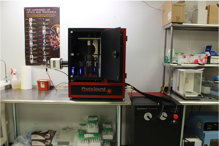
Fig 2. A small animal whole body in vivo imaging platform TriTom based on photoacoustic fluorescence tomography with an integrated PhotoSonus M laser system. Courtesy of PhotoSound Technologies, Inc.

ABSTRACT

This report highlights PhotoSonus M+, a tunable wavelength laser system recently introduced and developed specifically for photoacoustic imaging applications, where the highest imaging depth and resolution are required.

Read the full publication →

Citation

A. Juronis and M. Jašinskas, “Breakthrough instruments and products PhotoSonus M+ laser for photoacoustic imaging,” Review of Scientific Instruments, vol. 92, no. 5, p. 059502, 2021, doi: 10.1063/5.0053559.

PHOTOACOUSTIC TOMOGRAPHY TO ASSESSACUTE VASOACTIVITY OF SYSTEMIC VASCULATURE

Author(s): Huda, Kristieª; Lawrence, Dylanª; Lindsey, Sarah; Bayer, Carolynª*


ABSTRACT

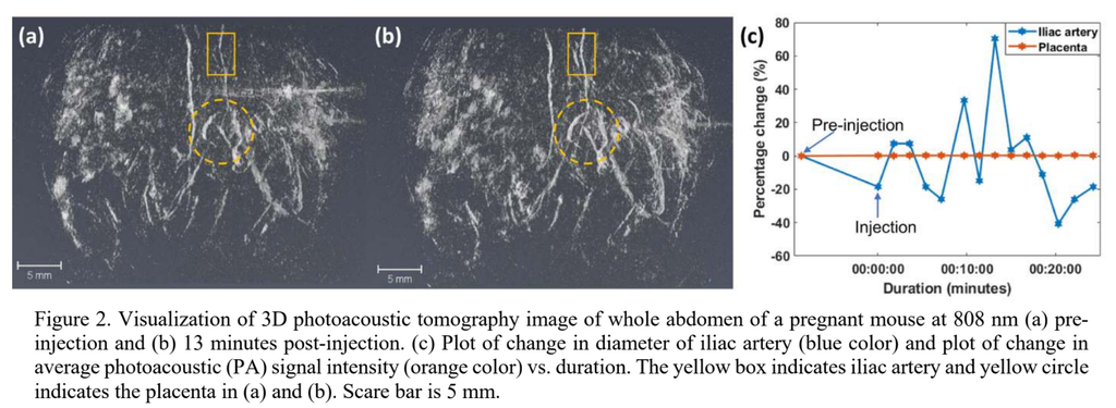
Vasoactivity is an important physiological indicator of cardiovascular health which is frequently measured using ex vivo vessels to determine functional mechanisms and evaluate pharmacological responses. Currently, there are no imaging methods available to assess vasoactivity in multiple vascular beds of living animals noninvasively. In this work, we have developed methods to use photoacoustic tomography to assess vasoactivity in vivo in systemic vasculature of living animals. A spherical-view photoacoustic tomography system was used to monitor acute vasodilation in the whole abdomen of a pregnant mouse in response to injection of G-1. After 3D image reconstruction, the diameter of the iliac artery and photoacoustic signal intensity of a placenta over time was measured. The artery and placenta had differential response to the vasodilator G-1. We validated the observed vasodilation of artery by monitoring the change in cross-sectional diameter of an individual artery using standard B-mode ultrasound imaging

Read the full publication →

Citation

K. Huda, D. Lawrence, S. Lindsey, and C. Bayer, Photoacoustic tomography to assess acute vasoactivity of systemic vasculature (SPIE BiOS). SPIE, 2022. https://doi.org/10.1117/12.2612093

PHOTOACOUSTIC IMAGING FOR IN VIVO QUANTIFICATION OF ALCOHOL-INDUCED STRUCTURAL AND FUNCTIONAL CHANGES IN CEREBRAL VASCULATURE IN HIGH ALCOHOL-PREFERRING MICE (HAP)

Authors: Augustine Meombe Mbolle, Hao Yang, Huabei Jiang*


ABSTRACT

Alcohol-induced structural and functional changes were studied in vivo by photoacoustic tomography (PAT) of the cerebrovascular system in selectively bred alcohol-preferring mice. High (HAP) and low (LAP) alcohol-preferring mice are replicate lines of mice selectively bred to prefer 10% (v/v) ethanol to water and water to ethanol, respectively, in a free-access two-bottle choice scenario. A cohort of 15 singly-housed alcohol-preferring mice (five HAP mice for the experimental group, five LAP mice for the control group, and five other LAP mice set aside) were given free-access two-bottle choice 10% ethanol (v/v) and water in 50-mL graduated drinking bottles mounted on each of their cages for 4 weeks prior to PAT brain scanning. A daily log of the volume of ethanol consumed over a 24-h period was kept. At the end of the fourth week, blood samples were collected from the HAP mice and blood ethanol concentrations (BECs) were measured to ascertain their levels of ethanol intoxication. The mice were then grouped into five weight-matched pairs of HAP and LAP for comparison purposes, and noninvasive in vivo PAT imaging was performed on each weight-matched pair. To mimic a binge drinking paradigm, mice were rearranged into four weight-matched groups of three animals each: an HAP mouse and two LAP mice. For each group, one HAP mouse and one LAP mouse received a 20% ethanol solution via intraperitoneal (i.p.) injection after 24 h of ethanol abstinence, in weight-based doses of 3 g/kg prior to imaging, while the last LAP mouse received a sham i.p. injection. PAT images of the brain were collected for 30 min thereafter. Cerebral vascular diameters for selected vessels of interest were extracted from the PAT images and compared between HAP mice and LAP mice. For the binge scenario, changes in vessel diameter and hemoglobin oxygen saturation were extracted from PAT images and studied over a 30-min duration. Vascular diameter was significantly smaller in HAP mice compared to LAP mice in weight-matched pairs. Hemoglobin-oxygen saturation and vessel diameter dropped more quickly in LAP mice than in HAP mice following a 20% ethanol i.p. injection (3 g/kg), with a 32% reduction in cerebrovascular diameter in a 30-min period. This study demonstrates the effectiveness of PAT in alcohol addiction imaging and diagnosis, and its feasibility in studying alcohol-induced changes in vascular structure and perfusion. It also adds to other bodies of evidence to suggest that the effects of binge drinking are more adverse in occasional drinkers than habitual drinkers.

Read the full publication →

Citation

A. Meombe Mbolle, H. Yang, and H. Jiang, “Photoacoustic Imaging for In Vivo Quantification of Alcohol-Induced Structural and Functional Changes in Cerebral Vasculature in High Alcohol-Preferring Mice (HAP),” Alcohol, vol. 100, pp. 23-30, 2022, doi: 10.1016/j.alcohol.2022.01.001.

TRANSFONTANELLE PHOTOACOUSTIC IMAGING: ULTRASOUND TRANSDUCER SELECTION ANALYSIS

Authors: Rayyan Manwar, Tarikul Islam, MD, Seyed Mohsen Ranjbaran, Kamran Avanaki


ABSTRACT

Transfontanelle ultrasound imaging (TFUI) is the conventional approach for diagnosing brain injury in neonates. Despite being the first stage imaging modality, TFUI lacks accuracy in determining the injury at an early stage due to degraded sensitivity and specificity. Therefore, a modality like photoacoustic imaging that combines the advantages of both acoustic and optical imaging can overcome the existing TFUI limitations. Even though a variety of transducers have been used in TFUI, it is essential to identify the transducer specification that is optimal for transfontanelle imaging using the photoacoustic technique. In this study, we evaluated the performance of 6 commercially available ultrasound transducer arrays to identify the optimal characteristics for transfontanelle photoacoustic imaging. We focused on commercially available linear and phased array transducer probes with center frequencies ranging from 2.5MHz to 8.5MHz which covers the entire spectrum of the transducer arrays used for brain imaging. The probes were tested on both in vitro and ex vivo brain tissue, and their performance in terms of transducer resolution, size, penetration depth, sensitivity, signal to noise ratio, signal amplification and reconstructed image quality were evaluated. The analysis of selected transducers in these areas allowed us to determine the optimal transducer for transfontanelle imaging, based on vasculature depth and blood density in tissue using ex vivo sheep brain. The outcome of this evaluation identified the two most suitable ultrasound transducer probes for transfontanelle photoacoustic imaging.

Read the full publication →

Citation

R. Manwar, M. T. Islam, S. M. Ranjbaran, and K. Avanaki, “Transfontanelle Photoacoustic Imaging: Ultrasound Transducer Selection Analysis,” Biomedical Optics Express, vol. 13, no. 2, pp. 676-693, 2022, doi: 10.1364/BOE.446087.

IN SITU X-RAY-INDUCED ACOUSTIC COMPUTED TOMOGRAPHY WITH A CONTRAST AGENT: A PROOF OF CONCEPT

Authors: Seongwook Choi,1; Sinyoung Park,1; Ayoung Pyo,2; Dong-Yeon Kim,; Jung-Joon Min,4 ; Changho Lee,4,5,6; Chulhong Kim,1,7


ABSTRACT

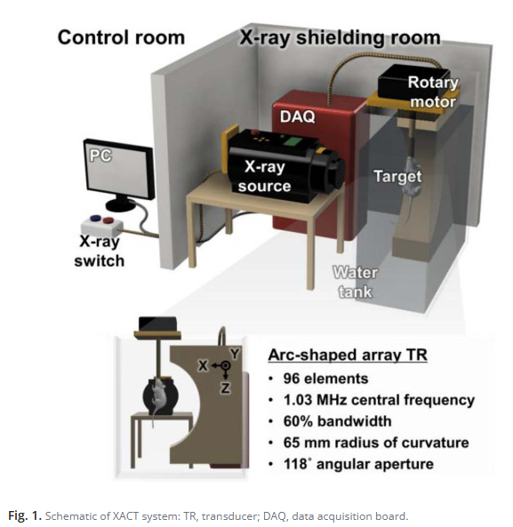
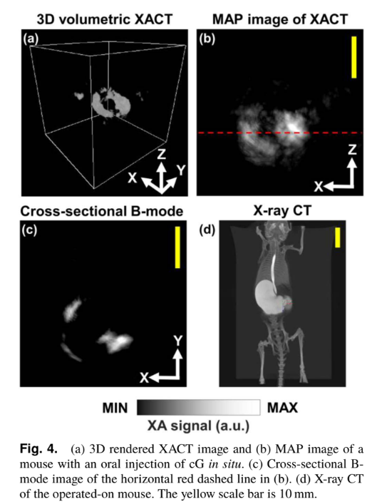
X-ray-induced acoustic computed tomography (XACT) has shown great potential as a hybrid imaging modality for real-time non-invasive x-ray dosimetry and low-dose three- dimensional (3D) imaging. While promising, one drawback of the XACT system is the underlying low signal-to-noise ratio (SNR), limiting its in vivo clinical use. In this Letter, we propose the first use of a conventional x-ray computed tomography contrast agent, Gastrografin, for improving the SNR of in situ XACT imaging. We obtained 3D volumetric XACT images of a mouse’s stomach with orally injected Gastrografin establishing the proposal’s feasibility. Thus, we believe, in the future, our proposed technique will allow in vivo imaging and expand or complement conventional x-ray modalities, such as radiotherapy and accelerators.

Read the full publication →

Citation

S. Choi, S. Park, A. Pyo, D.-Y. Kim, J.-J. Min, C. Lee, and C. Kim, “In situ x-ray-induced acoustic computed tomography with a contrast agent: a proof of concept,” Opt. Lett., vol. 47, no. 1, p. 90, 2022, doi: 10.1364/OL.447618.

SIZE-ADJUSTABLE RING-SHAPE PHOTOACOUSTIC TOMOGRAPHY IMAGER IN VIVO

Author(s): Daohuai Jiang1,2,3, Yifei Xu1, Hengrong Lan1,2,3, Yuting Shen1, Yifan Zhang1,Feng Gao1, Li Liu4*,  Fei Gao1,5*


ABSTRACT

Photoacoustic tomography (PAT) has become a novel biomedical imaging modality for scientific research and clinical diagnosis. It combines the advantages of spectroscopic optical absorption contrast and acoustic resolution with deep penetration. In this article, an imaging size-adjustable PAT system is proposed for potential clinical applications such as breast cancer detection and screening, which can adapt to imaging targets with various sizes. Comparing with the conventional PAT setup with a fixed radius ring shape ultrasound transducer (UT) array, the proposed system is more flexible for imaging diverse size targets based on sectorial ultrasound transducer arrays (SUTAs). Four SUTAs form a 128-channel UT array for photoacoustic detection, where each SUTA has 32 elements. Such four SUTAs are controlled by four stepper motors, respectively, and can change their distribution layout position to adapt for various imaging applications. In this proposed system, the radius of the imaging region of interest (ROI) can be adjusted from 50 to 100 mm, which is much more flexible than the conventional PAT system with a full ring UT array. The simulation experiments using the MATLAB k-wave toolbox demonstrate the feasibility of the proposed system. To further validate the proposed system, imaging of pencil leads made phantom, ex-vivo pork breast with indocyanine green (ICG) injected, and in-vivo human wrist, finger and ankle are conducted to prove its feasibility for potential clinical applications

Read the full publication →

Citation

D. Jiang, Y. Xu, H. Lan, Y. Shen, Y. Zhang, F. Gao, L. Liu, and F. Gao, "Size-Adjustable Ring-Shape Photoacoustic Tomography Imager In Vivo," Journal of Biophotonics, vol. 15, no. 7, 2022, doi: 10.1002/jbio.202200070.

NON-INVASIVE PHOTOACOUSTIC COMPUTED TOMOGRAPHY OF RAT HEART ANATOMY AND FUNCTION

Author(s): Li Lin, Xin Tong, Susana Cavallero, Yide Zhang, Shuai Na, Rui Cao, Tzung K. Hsiai, and Lihong V. Wang


ABSTRACT

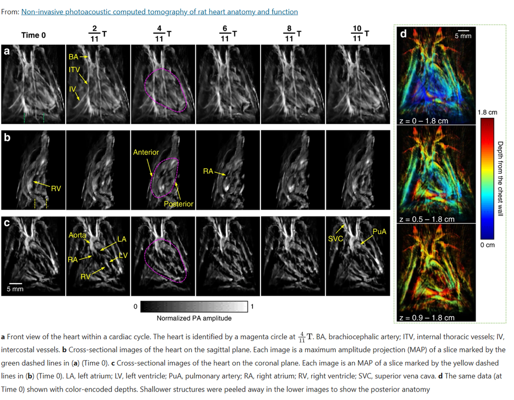
Photoacoustic computed tomography (PACT) combines optical contrast with acoustic-resolution imaging, but non-invasive cardiac imaging with this technique has been limited by signal loss and distortion caused by the chest wall. This work presents a 3D-PACT platform designed to overcome that limitation, using an optimized illumination and detection scheme to image rat hearts non-invasively — without surgical exposure of the chest — capturing a full cardiac scan in about 10 seconds. The system reveals both cardiac anatomy and intracardiac blood flow, gated to the animal’s ECG signal to freeze motion at each point in the cardiac cycle. Applied across healthy, hypertensive, and obese rat models, the technique distinguished differences in chamber size, wall thickness, and hemodynamics between groups, pointing toward potential future use in non-invasive, high-speed cardiac imaging of human neonates.

Read the full publication →

Citation

L. Lin, X. Tong, S. Cavallero, Y. Zhang, S. Na, R. Cao, T. K. Hsiai, and L. V. Wang, “Non-Invasive Photoacoustic Computed Tomography of Rat Heart Anatomy and Function,” Light: Science & Applications, vol. 12, 2023, doi: 10.1038/s41377-022-01053-7.

REAL-TIME, VOLUMETRIC IMAGING OF RADIATION DOSE DELIVERY DEEP INTO THE LIVER DURING CANCER TREATMENT

Authors: Wei Zhang, Ibrahim Oraiqat, Dale Litzenberg, Kai-Wei Chang, Scott Hadley, Noora Ba Sunbul, Martha M. Matuszak,  Christopher J. Tichacek, Eduardo G. Moros, Paul L. Carson, Kyle C. Cuneo, Xueding Wang, & Issam El Naqa


ABSTRACT

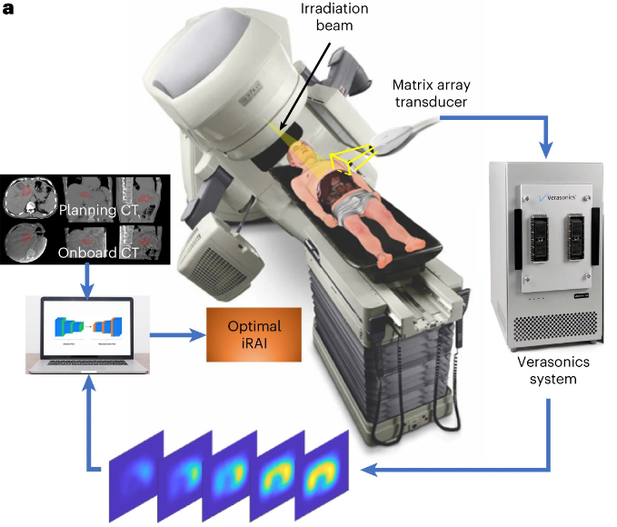
Ionizing radiation acoustic imaging (iRAI) allows online monitoring of radiation’s interactions with tissues during radiation therapy, providing real-time, adaptive feedback for cancer treatments. We describe an iRAI volumetric imaging system that enables mapping of the three-dimensional (3D) radiation dose distribution in a complex clinical radiotherapy treatment. The method relies on a two-dimensional matrix array transducer and a matching multi-channel preamplifier board. The feasibility of imaging temporal 3D dose accumulation was first validated in a tissue-mimicking phantom. Next, semiquantitative iRAI relative dose measurements were verified in vivo in a rabbit model. Finally, real-time visualization of the 3D radiation dose delivered to a patient with liver metastases was accomplished with a clinical linear accelerator. These studies demonstrate the potential of iRAI to monitor and quantify the 3D radiation dose deposition during treatment, potentially improving radiotherapy treatment efficacy using real-time adaptive treatment.

Read the full publication →

Citation

W. Zhang, I. Oraiqat, D. Litzenberg, K.-W. Chang, S. Hadley, N. Ba Sunbul, M. M. Matuszak, C. J. Tichacek, E. G. Moros, P. L. Carson, K. C. Cuneo, X. Wang, and I. El Naqa, "Real-Time, Volumetric Imaging of Radiation Dose Delivery Deep into the Liver during Cancer Treatment," Nature Biotechnology, 2023, doi: 10.1038/s41587-022-01593-8.