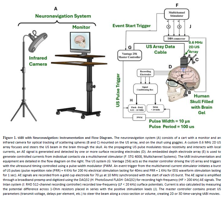
Author(s): Nadia Abu Farha1, Parker Harris, Margaret Allard, Teodoro Trujillo, Stephen Cowen, Katalin Gothard4, Martin Weinand, Paul Larson, Ying-Hui Chou, Nan-Kuei Chen, Yeonjoon Cheong, Matthew O’Donnell6, Russell S Witte
N. Abu Farha, P. Harris, M. Allard, T. Trujillo, S. Cowen, K. Gothard, M. Weinand, P. Larson, Y.-H. Chou, N.-K. Chen, Y. Cheong, M. O'Donnell, and R. S. Witte, "Transcranial acoustoelectric imaging (tABI) of seizure activity in human head model with neuronavigation," Journal of Neural Engineering, vol. 23, no. 3, art. 036038, 2026, doi: 10.1088/1741-2552/ae7764.
Author(s): K. C. Cuneo, W. Zhang, D. Litzenberg, Y. Huang, S. Hadley, I. Oraiqat, K.-W. Chang, G. Gonzalez, S. Dykstra, M. Zhang, E. G. Moros, P. L. Carson, I. El Naqa, and X. Wang
ABSTRACT
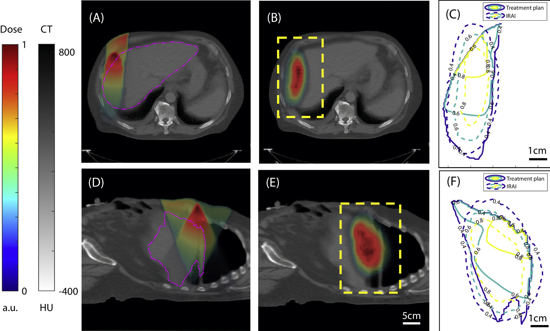
This study aimed to evaluate the feasibility of using ionizing radiation acoustic imaging (iRAI) to map the delivered dose in patients receiving radiation therapy (RT) with various treatment techniques, including 3-dimensional conformal RT, intensity modulated RT, and volumetric modulated arc therapy.
Patients with intra-abdominal cancer were enrolled in a prospective clinical trial after providing informed consent. Patients were treated with stereotactic body RT using standard clinical techniques with the addition of volumetric iRAI for real-time mapping of 3-dimensional radiation dose deposition.
The minimal detectable dose was approximately 10 cGy. The overall shape of the dose distribution and the location of dose deposition, as determined by iRAI, matched the corresponding treatment plan. A gamma passing rate of 75.97% ± 11.12% and 90.99% ± 6.61% with a 10 mm/10% distance to agreement/dose difference suggests good agreement between the iRAI measurement and the treatment plan in both the liver volume and the planning target volume under the current system resolution. A structural similarity value of 0.6284 ± 0.1678 demonstrates that the iRAI measurements have shown structural pattern agreement with the treatment plan within the planning target volume.
This study demonstrates the clinical feasibility of iRAI to monitor radiation dose delivery to deep targets in real time during treatment. This information can be integrated into treatment delivery systems to improve safety and facilitate the adaptation of RT. Despite the promising results achieved with the current form of technology, limitations in detection sensitivity, reconstruction accuracy, and acoustic coupling still need to be addressed.
K. C. Cuneo, W. Zhang, D. Litzenberg, Y. Huang, S. Hadley, I. Oraiqat, K.-W. Chang, G. Gonzalez, S. Dykstra, M. Zhang, E. G. Moros, P. L. Carson, I. El Naqa, and X. Wang, "Clinical Pilot Study of Ionizing Radiation Acoustic Imaging for Real-Time Visualization of Radiation Therapy Dose Delivery in Cancer Patients," Int. J. Radiat. Oncol. Biol. Phys., vol. 125, no. 3, pp. 959-969, 2026, doi: 10.1016/j.ijrobp.2025.12.006.
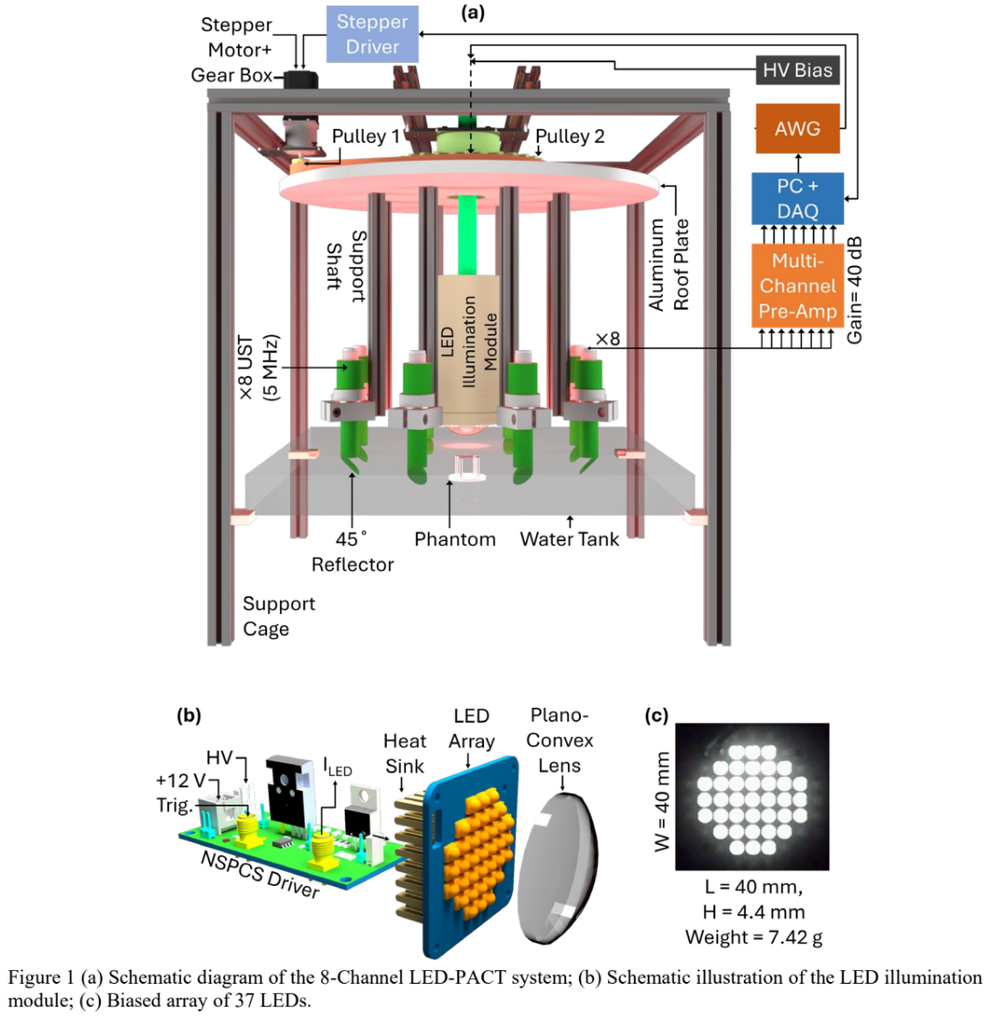
Light-emitting diodes (LEDs) emerge as a viable alternative to pulsed lasers in photoacoustic computed tomography (PACT) due to their low cost, compact form factor, wavelength flexibility, and reduced safety concerns. Unlike class-IV laser systems, LED-based excitation eliminates regulatory constraints while enabling high pulse-repetition rates (PRRs) and flexible modulation for improved signal averaging and contrast. We present a high-speed nanosecond pulsed LEDbased PACT (LED-PACT) system optimized for rapid tomographic imaging using multiple single-element ultrasound transducers (USTs) arranged in a circular geometry. A custom nanosecond pulsed current source (NSPCS) drives 37 infrared LEDs at 850 nm, delivering ~0.19 mJ per pulse. Sub-100 ns excitation enables PRRs ≥10 kHz, allowing fast data acquisition with reduced motion artifacts. Imaging speed is further increased by employing eight single-element USTs that rotate mechanically around the target. This configuration reduces the effective angular step size to 360˚/n, where n is the number of transducers, achieving substantially faster full-angle coverage than conventional single-UST systems. The system performance was evaluated using absorbing phantoms. Delay-and-sum (DAS) reconstructed images demonstrate clear visualization of target geometry at scan times as short as 0.25 s, corresponding to a frame rate of 4 Hz. These results confirm that the combined use of high-PRR nanosecond LED excitation and multi-UST acquisition enables sub-second PACT imaging while maintaining stable signal generation and sufficient image contrast. The proposed architecture provides a compact, scalable, and cost-effective pathway toward high-speed PACT systems for dynamic biomedical imaging applications.
A. Das and M. Pramanik, Nanosecond Pulsed Light Emitting Diode (LED) Based High-Speed Photoacoustic Computed Tomography Using Multiple Single-Element Ultrasound Transducers (SPIE BiOS). SPIE, 2026. doi: 10.1117/12.3079921.
Author(s): Aixa Aguilera-Garrido, Marie Millard, Mathilde Bouché, Pascal Lemière, Nadia Canilho, Andreea Pasc, Kazunori Kataoka, Joachim F.R. Van Guyse, Henri-Pierre Lassalle, Yann Bernhard
ABSTRACT
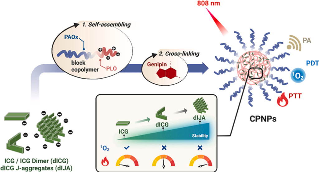
Photothermal therapy (PTT) offers a minimally invasive approach for treating hypoxic tumors, while photodynamic therapy (PDT) is limited by its oxygen dependence. Indocyanine green (ICG) is a clinically approved near-infrared (NIR) dye with potential for PTT and PDT, but its application is hindered by poor photostability, rapid clearance, and aggregation, resulting in unfavorable pharmacokinetics and limited phototherapeutic performances. Here, we address these limitations through a combination of chemical transformation of ICG by dimerization, controlled supramolecular assembly into dye aggregates, and a tailored formulation strategy. We systematically compared three forms, monomeric ICG, its covalent dimer (dICG), and the corresponding J-aggregate (dIJA), each formulated into polyion complex nanoparticles (PNPs) using poly(2-ethyl-2-oxazoline)-b-poly(L-ornithine) as the polyelectrolyte carrier matrix. Subsequent genipin-mediated core crosslinking produced crosslinked nanoparticles (CPNPs) with enhanced stability, high encapsulation efficiency, and preserved optical characteristics. CPNPs consistently outperformed free dye and non-crosslinked PNPs, showing improved colloidal stability, enhanced photostability, and higher photothermal performances. In FaDu cells, ICG-loaded CPNPs mediated oxygen-dependent photodynamic effects, whereas dICG- and dIJA-loaded CPNPs induced oxygen-independent photothermal cytotoxicity under NIR irradiation. Our integrated comparison demonstrates that controlling the molecular form and supramolecular assembly of ICG derivatives enables tunable NIR phototherapy, identifying dICG- and dIJA-based CPNPs as promising platforms for the treatment of hypoxic tumors by photoacoustic imaging (PAI) guided PTT.
A. Aguilera-Garrido, M. Millard, M. Bouché, P. Lemière, N. Canilho, A. Pasc, K. Kataoka, J. F. R. Van Guyse, H.-P. Lassalle, and Y. Bernhard, "Phototherapy mode control by tuning molecular and supramolecular forms of indocyanine green in crosslinked polyion complex nanoparticles," Materials Today Advances, vol. 31, p. 100891, 2026, doi: 10.1016/j.mtadv.2026.100891.
Author(s): A. M. Yu, X. Huang, S. M. A. Morais, J. H. Park, D. A. Zopf, S. J. Hollister, and S. Emelianov
ABSTRACT
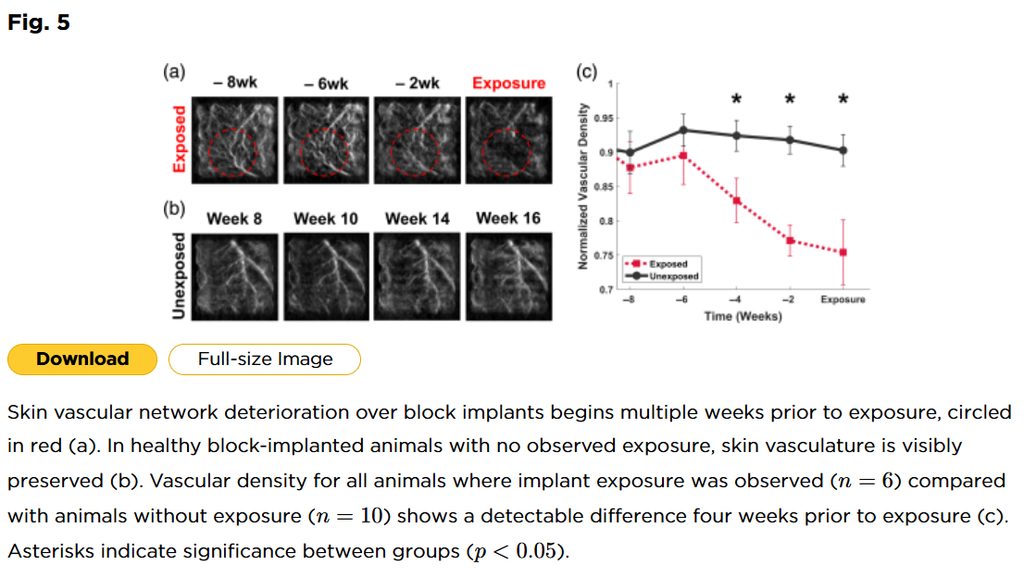
Craniofacial implants are prone to skin necrosis and exposure, likely due to implant-induced stress and ischemia. However, this relationship has not been quantitatively validated. Identifying an imaging biomarker of skin vascular health could confirm this mechanism and help predict skin failure to mitigate implant complications.
Photoacoustic tomography (PAT) was used to monitor changes in the skin vasculature surrounding subcutaneous implants with the goal of obtaining a quantitative metric predictive of implant exposure.
Three designs of 3D-printed porous polycaprolactone (PCL) constructs—unimodal block, bimodal block, and unimodal dome—were implanted in 16 hairless mice. PAT was performed biweekly for 16 weeks, and a skeletonization algorithm was applied to quantify vascular density in skin overlying the implants.
Mice that developed implant exposure (N=6) exhibited a progressive decline in vascular density beginning 6 weeks before visible exposure, whereas nonexposed mice (N=10) remained stable. Group differences were significant 4 weeks (p=0.031) and 2 weeks (p=0.001) before exposure onset.
These findings establish a quantitative temporal relationship between vascular ischemia and implant exposure. PAT-derived vascular density serves as a predictive biomarker of skin failure, which can be used to enable interventional treatment and improve implant designs.
A. M. Yu, X. Huang, S. M. A. Morais, J. H. Park, D. A. Zopf, S. J. Hollister, and S. Emelianov, "Quantitative photoacoustic tomography of skin vasculature predicts subcutaneous implant exposure," J. Biomed. Opt., vol. 31, no. 2, p. 026005, 2026, doi: 10.1117/1.JBO.31.2.026005.
Author(s): M. Pérez-Liva, M. Alonso de Leciñana, M. Gutiérrez-Fernández, J. Camacho Sosa Dias, J. F. Cruza, J. Rodríguez-Pardo, I. García-Suárez, F. Laso-García, J. L. Herraiz, and L. Elvira Segura
ABSTRACT
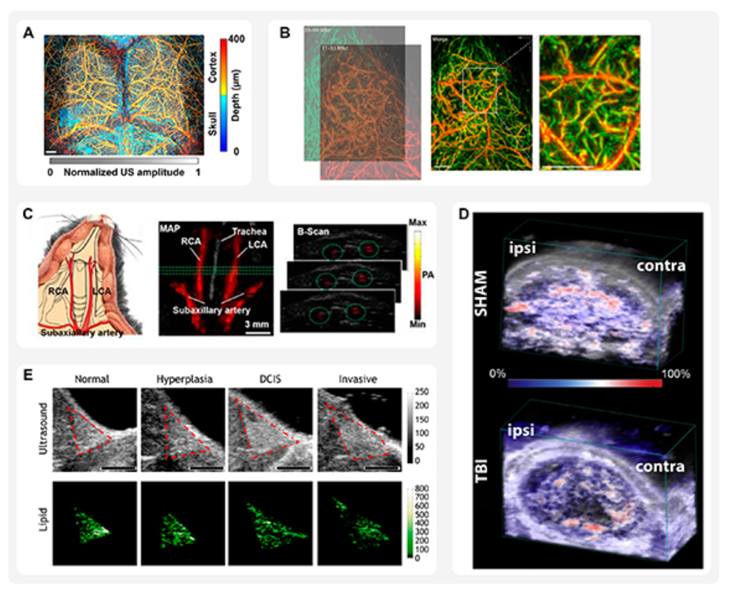
Photoacoustic (PA) imaging, by integrating optical and ultrasound (US) modalities, combines high spatial resolution with deep tissue penetration, making it a transformative tool in biomedical research. This review presents a comprehensive analysis of the current status of dual PA/US imaging technologies, emphasising their applications in preclinical research. It details advancements in light excitation strategies, including tomographic and microscopic modalities, innovations in pulsed laser and alternative light sources, and US instrumentation. The review further explores preclinical methodologies, encompassing dedicated instrumentation, signal processing, and data analysis techniques essential for PA/US systems. Key applications discussed include the visualisation of blood vessels, micro-circulation, and tissue perfusion; diagnosis and monitoring of inflammation; evaluation of infections, atherosclerosis, burn injuries, healing, and scar formation; assessment of liver and renal diseases; monitoring of epilepsy and neurodegenerative conditions; studies on brain disorders and preeclampsia; cell therapy monitoring; and tumour detection, staging, and recurrence monitoring. Challenges related to imaging depth, resolution, cost, and the translation of contrast agents to clinical practice are analysed, alongside advancements in high-speed acquisition, artificial intelligence-driven reconstruction, and innovative light-delivery methods. While clinical translation remains complex, this review underscores the crucial role of preclinical studies in unravelling fundamental biomedical questions and assessing novel imaging strategies. Ultimately, this review delves into the future trends of dual PA/US imaging, highlighting its potential to bridge preclinical discoveries with clinical applications and drive advances in diagnostics, therapeutic monitoring, and personalised medicine.
M. Pérez-Liva, M. Alonso de Leciñana, M. Gutiérrez-Fernández, J. Camacho Sosa Dias, J. F. Cruza, J. Rodríguez-Pardo, I. García-Suárez, F. Laso-García, J. L. Herraiz, and L. Elvira Segura, "Dual photoacoustic/ultrasound technologies for preclinical research: current status and future trends," Physics in Medicine & Biology, vol. 70, art. 07TR01, 2025, doi: 10.1088/1361-6560/adb368.
Author(s): Tri Vu, Paul Klippel, Aidan J. Canning, Chenshuo Ma, Huijuan Zhang, Ludmila A. Kasatkina, Yuqi Tang, Jun Xia, Vladislav V. Verkhusha, Tuan Vo-Dinh, Yun Jing, Junjie Yao
ABSTRACT
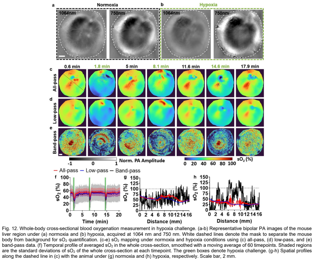
In photoacoustic computed tomography (PACT) with short-pulsed laser excitation, wideband acoustic signals are generated in biological tissues with frequencies related to the effective shapes and sizes of the optically absorbing targets. Low-frequency photoacoustic signal components correspond to slowly varying spatial features and are often omitted during imaging due to the limited detection bandwidth of the ultrasound transducer, or during image reconstruction as undesired background that degrades image contrast. Here we demonstrate that low-frequency photoacoustic signals, in fact, contain functional and molecular information, and can be used to enhance structural visibility, improve quantitative accuracy, and reduce spare-sampling artifacts. We provide an in-depth theoretical analysis of low-frequency signals in PACT, and experimentally evaluate their impact on several representative PACT applications, such as mapping temperature in photothermal treatment, measuring blood oxygenation in a hypoxia challenge, and detecting photoswitchable molecular probes in deep organs. Our results strongly suggest that low-frequency signals are important for functional and molecular PACT.
T. Vu, P. Klippel, A. J. Canning, C. Ma, H. Zhang, L. A. Kasatkina, Y. Tang, J. Xia, V. V. Verkhusha, T. Vo-Dinh, Y. Jing, and J. Yao, “On the importance of low-frequency signals in functional and molecular photoacoustic computed tomography,” IEEE Trans. Med. Imaging, vol. 43, no. 2, pp. 771-783, 2024, doi: 10.1109/TMI.2023.3320668.
Author(s):Vinoin Devpaul Vincely, Xingjian Zhong, Kristie Huda, Swathi P. Katakam, Joshua C. Kays, Allison M. Dennis, Carolyn L. Bayer
ABSTRACT
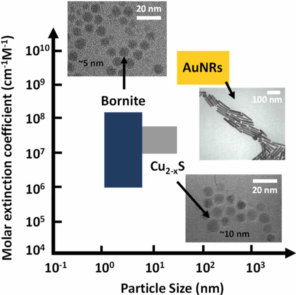
In this study, we demonstrate the potential of the bornite crystal structure (Cu5FeS4) of copper iron sulfide as a second near infrared (NIR-II) photoacoustic (PA) contrast agent. Bornite exhibits comparable dose-dependent biocompatibility to copper sulfide nanoparticles in a cell viability study with HepG2 cells, while exhibiting a 10-fold increase in PA amplitude. In comparison to other benchmark contrast agents at similar mass concentrations, bornite demonstrated a 10× increase in PA amplitude compared to indocyanine green (ICG) and a 5× increase compared to gold nanorods (AuNRs). PA signal was detectable with a light pathlength greater than 5 cm in porcine tissue phantoms at bornite concentrations where in vitro cell viability was maintained. In vivo imaging of mice vasculature resulted in a 2× increase in PA amplitude compared to AuNRs. In summary, bornite is a promising NIR-II contrast agent for deep tissue PA imaging.
V. D. Vincely, X. Zhong, K. Huda, S. P. Katakam, J. C. Kays, A. M. Dennis, and C. L. Bayer, "Bornite (Cu5FeS4) nanocrystals as an ultrasmall biocompatible NIR-II contrast agent for photoacoustic imaging," Photoacoustics, vol. 40, p. 100649, 2024, doi: 10.1016/j.pacs.2024.100649.
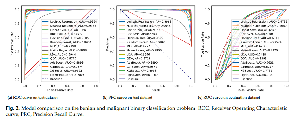
Colorectal cancer is a deadly disease that has become increasingly prevalent in recent years. Early detection is crucial for saving lives, but traditional diagnostic methods such as colonoscopy and biopsy have limitations. Colonoscopy cannot provide detailed information within the tissues affected by cancer, while biopsy involves tissue removal, which can be painful and invasive. In order to improve diagnostic efficiency and reduce patient suffering, we studied machine-learningbased approach for colorectal tissue classification that uses acoustic resolution photoacoustic microscopy (ARPAM). With this tool, we were able to classify benign and malignant tissue using multiple machine learning methods. Our results were analyzed both quantitatively and qualitatively to evaluate the effectiveness of our approach.
S. Tong, P. Ge, Y. Jiao, Z. Ma, Z. Li, L. Liu, F. Gao, X. Du, and F. Gao, "Machine-Learning-Based Colorectal Tissue Classification via Acoustic Resolution Photoacoustic Microscopy," arXiv, 2023, doi: 10.48550/arXiv.2307.08556.
Author(s): Zohar Or, Ahiad R. Levi, Yoav Hazan and Amir Rosenthal
ABSTRACT
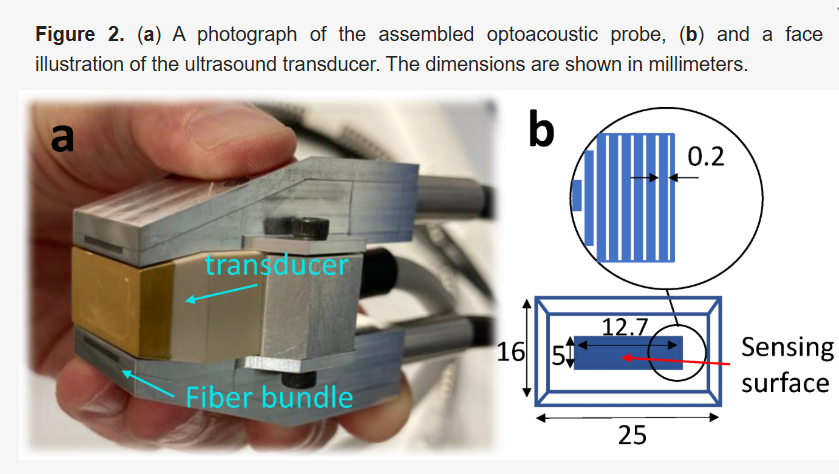
he ability to rapidly locate blood vessels in patients is important in many clinical applications, e.g., in catheterization procedures. Optical techniques, including visual inspection, generally suffer from a reduced performance at depths below 1 mm, while ultrasound and optoacoustic tomography are better suited to a typical depth on the scale of 1 cm and require an additional spacer between the tissue and transducer in order to image the superficial structures at the focus plane. For this work, we developed a hand-held optoacoustic probe, designed for localizing blood vessels from the contact point down to a depth of 1 cm, without the use of a spacer. The probe employs a flat lens-free ultrasound array, enabling a largely depth-independent response down to a depth of 1 cm, at the expense of low elevational resolution. Specifically, while in lens-based probes, the acoustic signals from outside the focal region suffer from distortion, in our probe, only the amplitude of the signal varies with depth, thus leading to an imaging quality that is largely depth-independent in the imaged region. To facilitate miniaturization, dark-field illumination is used, whereby light scattering from the tissue is exploited to homogenize the sensitivity field.
Z. Or, A. R. Levi, Y. Hazan, and A. Rosenthal, "Hand-Held Optoacoustic System for the Localization of Mid-Depth Blood Vessels," Photonics, vol. 9, no. 12, art. 907, 2022, doi: 10.3390/photonics9120907.
Manage Consent
To provide the best experiences, we use technologies like cookies to store and/or access device information. Consenting to these technologies will allow us to process data such as browsing behavior or unique IDs on this site. Not consenting or withdrawing consent, may adversely affect certain features and functions.
Functional
Always active
The technical storage or access is strictly necessary for the legitimate purpose of enabling the use of a specific service explicitly requested by the subscriber or user, or for the sole purpose of carrying out the transmission of a communication over an electronic communications network.
Preferences
The technical storage or access is necessary for the legitimate purpose of storing preferences that are not requested by the subscriber or user.
Statistics
The technical storage or access that is used exclusively for statistical purposes.The technical storage or access that is used exclusively for anonymous statistical purposes. Without a subpoena, voluntary compliance on the part of your Internet Service Provider, or additional records from a third party, information stored or retrieved for this purpose alone cannot usually be used to identify you.
Marketing
The technical storage or access is required to create user profiles to send advertising, or to track the user on a website or across several websites for similar marketing purposes.