Recent Posts

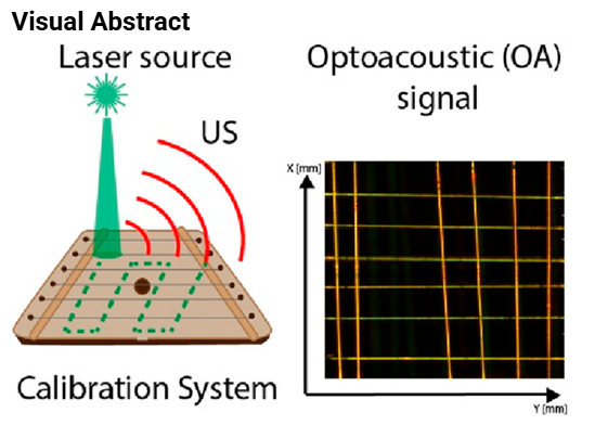
Carbon Nanotube Microscale Fiber Grid as an Advanced Calibration System for Multispectral Optoacoustic Imaging

Author(s): Margarita R. Chetyrkina, Julijana Cvjetinovic, Fedor S. Fedorov, Stanislav V. Perevoschikov, Ekaterina S., Prikhozhdenko,Bjørn F. Mikladal, Yuri G. Gladush, Albert G. Nasibulin,Dmitry A. Gorin

ABSTRACT

Optoacoustic (photoacoustic) imaging has gained tremendous attention in research and in clinical practice as a point-of-care system for noninvasive, fast, and safe tests. The first optoacoustic (OA) tomograph has recently passed the Food and Drug Administration (FDA) approval stage for clinical applications aimed at early breast cancer diagnostics. Furthermore, a broad application of OA imaging for Biomedical and Materials Science fields requires a proper tool to test the equipment and verify the quality of the measurements on a daily basis. In the present work, we propose fibers based on single-walled carbon nanotubes (SWCNTs) as a material for designing a stable and reliable calibration grid. The main advantage of the developed test system is the broad optical absorption of SWCNT-based fibers, ranging from visible to mid-infrared regions. Inspired by stringed instruments, we elaborate a grid to calibrate and verify spatial resolution in three projections and sensitivity of OA imaging systems. Thus, the real calibration grid parameters, such as fiber length and diameter, could be translated to the OA signal measurements. This proof-of-the-concept study evaluates the geometry of fibers, that is, the length/diameter and design of fibers, such as free-standing/twisted, and shows the fabrication procedure of the calibration grid prototype toward the successful validation of the OA imaging system, including raster-scanning optoacoustic mesoscopy (RSOM) at one wavelength and tomography at several wavelengths, which have grand prospects in preclinical and clinical practices. Besides, the more advanced geometry based on double-twisted fibers, or twistrons, applied here provided us with a chance to reach the lower resolution limit for RSOM because of the difference in diameter between the thin and thick parts in the morphology is verified by scanning electron microscopy.

Read the full publication →

Citation

M. R. Chetyrkina, J. Cvjetinovic, F. S. Fedorov, S. V. Perevoschikov, E. S. Prikhozhdenko, B. F. Mikladal, Y. G. Gladush, A. G. Nasibulin, and D. A. Gorin, "Carbon Nanotube Microscale Fiber Grid as an Advanced Calibration System for Multispectral Optoacoustic Imaging," ACS Photonics, vol. 9, no. 10, pp. 3429-3439, 2022/10/19 2022, doi: 10.1021/acsphotonics.2c01074.
Targeted contrast agents and activatable probes for photoacoustic imaging of cancer

Author(s): Zhenxiang Zhao, Chelsea Swartchick, Jefferson Chan


ABSTRACT

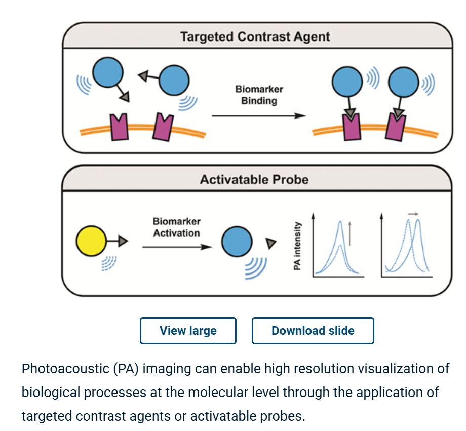
Photoacoustic (PA) imaging has emerged as a powerful technique for the high resolution visualization of biological processes within deep tissue. Through the development and application of exogenous targeted contrast agents and activatable probes that can respond to a given cancer biomarker, researchers can image molecular events in vivo during cancer progression. This information can provide valuable details that can facilitate cancer diagnosis and therapy monitoring. In this tutorial review, we provide a step-by-step guide to select a cancer biomarker and subsequent approaches to design imaging agents for in vivo use. We envision this information will be a useful summary to those in the field, new members to the community, and graduate students taking advanced imaging coursework. We also highlight notable examples from the recent literature, with emphasis on the molecular designs and their in vivo PA imaging performance. To conclude, we provide our outlook and future perspective in this exciting field.

Read the full publication →

Citation

Z. Zhao, C. B. Swartchick, and J. Chan, "Targeted contrast agents and activatable probes for photoacoustic imaging of cancer," Chem Soc Rev, vol. 51, no. 3, pp. 829-868, Feb 7 2022, doi: 10.1039/d0cs00771d.

Assessment of a Single-Element Scanning System for Enhanced Photoacoustic Imaging of Brain Hemorrhage

Author(s): Juliana Benavides Lara, Ravi Prakash, Kamran Avanaki


ABSTRACT

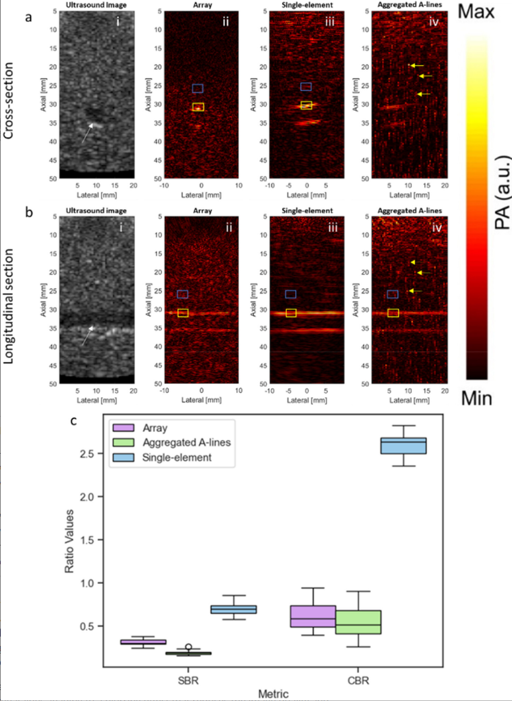
The use of photoacoustic brain imaging for hemorrhage detection holds significant clinical importance. This study focuses on the performance of sensitivity and detection capabilities of a single-element scanning system, considering the remarkable signal-to-noise ratio of photoacoustic signals generated by a single-element transducer. By employing blood vessel-like phantoms and ex vivo brain phantoms, we demonstrated the superior efficacy of the single-element scanning method over the transducer array system in the context of brain hemorrhage detection. This research highlights the potential for enhancing hemorrhage detection sensitivity through careful design and optimization of the proposed method, thereby increasing its viability for clinical application.

Read the full publication →

Citation

J. Benavides Lara, R. Prakash, and K. Avanaki, "Assessment of a Single-Element Scanning System for Enhanced Photoacoustic Imaging of Brain Hemorrhage," Journal of Biophotonics, vol. 18, no. 3, art. e202400153, 2025, doi: 10.1002/jbio.202400153.

High-throughput photoacoustic tomography by integrated robotics and automation

Author(s): N. Marshall, H.-P. Brecht, W. Thompson, D. Lawrence, V. Marshall, S. Toler, S. Emelianov, A. Yu, M. A. Anastasio, U. Villa, J. Maxwell, and S. Ermilov


ABSTRACT

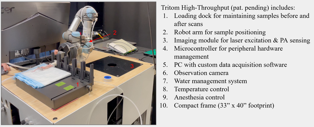
We present the development of a photoacoustic tomography (PAT) imaging system with the demonstrated capability of obtaining high-throughput scans at a sustained rate of under 1 minute per animal using integrated robotics to assist in 3D PAT collection. This is a considerable achievement as there is currently no existing commercial or research PAT whole-body imaging system capable of high-throughput applications (15-20 animals per hour). High-throughput experimentation is imperative in the development, characterization, and use of rodent models of human diseases as it increases the number of animals that can be evaluated within a single experiment and may reduce the time under anesthesia for each animal, thereby improving the stability, duration, and confidence of longitudinal studies The developed system features coordinated automation for robotic animal manipulation, anesthesia distribution, temperature regulation, water management, laser excitation, and photoacoustic detection. Furthermore, as shown in validation studies using phantoms and live murine models, the prototype imaging platform demonstrates high-throughput performance while retaining high sensitivity and high resolution.

Read the full publication →

Citation

N. Marshall, H.-P. Brecht, W. Thompson, D. Lawrence, V. Marshall, S. Toler, S. Emelianov, A. Yu, M. A. Anastasio, U. Villa, J. Maxwell, and S. Ermilov, High-throughput photoacoustic tomography by integrated robotics and automation (SPIE BiOS). SPIE, 2024. doi: 10.1117/12.3005569.

In vivo noninvasive systemic myography of acute systemic vasoactivity in female pregnant mice

Author(s): Kristie Huda, Dylan J. Lawrence, Weylan Thompson, Sarah H. Lindsey & Carolyn L. Bayer


ABSTRACT

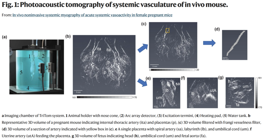
Altered vasoactivity is a major characteristic of cardiovascular and oncological diseases, and many therapies are therefore targeted to the vasculature. Therapeutics which are selective for the diseased vasculature are ideal, but whole-body selectivity of a therapeutic is challenging to assess in practice. Vessel myography is used to determine the functional mechanisms and evaluate pharmacological responses of vascularly-targeted therapeutics. However, myography can only be performed on ex vivo sections of individual arteries. We have developed methods for implementation of spherical-view photoacoustic tomography for non-invasive and in vivo myography. Using photoacoustic tomography, we demonstrate the measurement of acute vascular reactivity in the systemic vasculature and the placenta of female pregnant mice in response to two vasodilators. Photoacoustic tomography simultaneously captures the significant acute vasodilation of major arteries and detects selective vasoactivity of the maternal-fetal vasculature. Photoacoustic tomography has the potential to provide invaluable preclinical information on vascular response that cannot be obtained by other established methods.

Read the full publication →

Citation

Kristie Huda, Dylan J. Lawrence, Weylan Thompson, and Carolyn L. Bayer, "In vivo noninvasive systemic myography of acute systemic vasoactivity in female pregnant mice", Nature Communications, vol. 14, 2023. https://doi.org/10.1038/s41467-023-42041-8

Radiomics-driven perfusion prediction in clinical photoacoustic foot imaging

Author(s): Chuqin Huang a, Yanda Cheng a, Xiaoyu Zhang b, Ye Zhan c, Wenhan Zheng a, Isabel Komornicki d, Linda M. Harris d, Wenyao Xu b, Jun Xia a


ABSTRACT

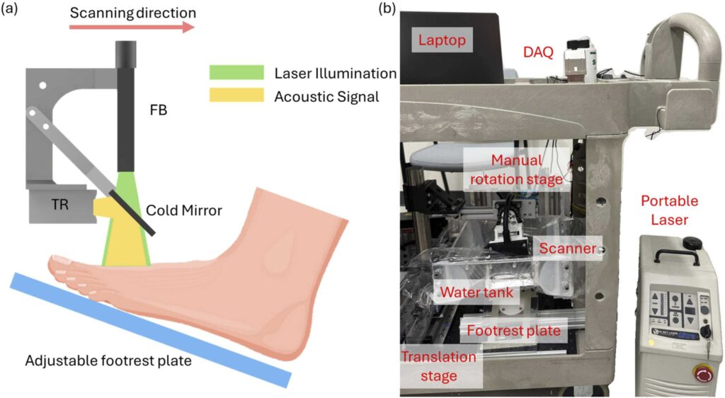
Accurate assessment of tissue perfusion is essential for managing chronic foot ulcers in patients with diabetes and peripheral arterial disease. While photoacoustic (PA) imaging enables high-resolution visualization of vascular structures, current perfusion evaluation methods are limited. We propose a fully automated radiomics-based framework for predicting perfusion conditions using single-wavelength clinical PA foot imaging. Radiomics features were extracted from both raw radiofrequency (RF) signals and reconstructed maximum amplitude projection (MAP) images. After reproducibility testing and statistical filtering, features were ranked using a combined minimum redundancy maximum relevance (mRMR) and ReliefF approach. A k-nearest neighbors ensemble model trained on eight selected features achieved an area under the curve (AUC) of 0.90 (training) and 0.94 (test). The selected features corresponded with physiological indicators such as vessel density, tissue structure, and vascular discontinuity. This study demonstrates a reliable and interpretable method for perfusion assessment in PA imaging with strong clinical potential.

Read the full publication →

Citation

C. Huang, Y. Cheng, X. Zhang, Y. Zhan, W. Zheng, I. Komornicki, L. M. Harris, W. Xu, and J. Xia, "Radiomics-driven perfusion prediction in clinical photoacoustic foot imaging," Photoacoustics, vol. 46, art. 100776, 2025, doi: 10.1016/j.pacs.2025.100776.

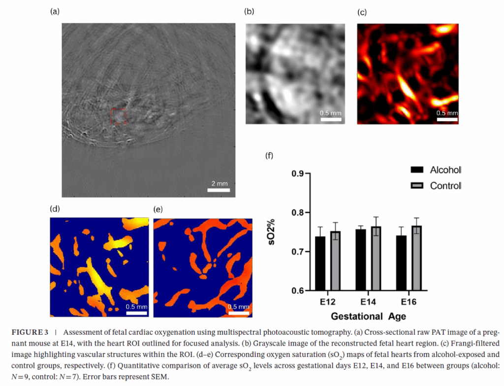
Investigating the Impact of Prenatal Alcohol Exposure on Fetal Cardiac Development Using Photoacoustic Tomography

Author(s): M. F. Tanvir, H. Yang, J. D. Gardner, and H. Jiang


ABSTRACT

Prenatal alcohol exposure (PAE) is a leading cause of developmental abnormalities, yet its effects on fetal cardiac development remain understudied. We employed real-time, label-freemultispectral photoacoustic tomography (PAT) to noninvasively assess cardiac development in mouse fetuses exposed to chronic alcohol. Using a custom-built PAT system, fetal hearts were imaged from E12 to E16 in alcohol-exposed (3 g/kg ethanol via oral gavage, n = 9) and control (n = 7) CD-1 mice. PAT enabled quantitative measurements of cardiac morphology, oxygen saturation (sO2), and heart rate. Alcohol-exposed fetuses exhibited consistently lower sO2 and greater heart rate variability, particularly at later gestational stages. While structural growth progressed in both groups, functional impairments became more pronounced with alcohol exposure. These findings suggest PAE alters fetal cardiovascular regulation despite normal anatomical development. This study highlights the utility of PAT as a high-resolution, noninvasive tool for monitoring fetal cardiac health and supports its potential application in developmental biology and prenatal diagnostics.

Read the full publication →

Citation

M. F. Tanvir, H. Yang, J. D. Gardner, and H. Jiang, "Investigating the Impact of Prenatal Alcohol Exposure on Fetal Cardiac Development Using Photoacoustic Tomography," Journal of Biophotonics, art. e202500215, 2025, doi: 10.1002/jbio.202500215.

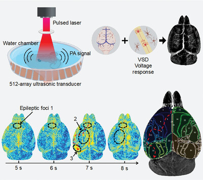
Direct Monitoring of Whole-Brain Electrodynamics via High-Spatiotemporal-Resolution Photoacoustics with Voltage-Sensitive Dye

Author(s): W. Pang, B. Zhu, H. Li, Y. Zhou, C. M. Woo, X. Huang, T. Zhong, H. Lo, L. Wang, P. Lai, and L. Nie


ABSTRACT

This work proposes a novel method for monitoring deep-brain electrodynamics with a voltage-sensitive dye (VSD)-based photoacoustic imaging platform, called PA-VSD. This platform is demonstrated to allow for effectively localizing epileptic foci and visualizing voltage conduction pathways in a high spatiotemporal resolution.

Read the full publication →

Citation

W. Pang, B. Zhu, H. Li, Y. Zhou, C. M. Woo, X. Huang, T. Zhong, H. Lo, L. Wang, P. Lai, and L. Nie, "Direct Monitoring of Whole-Brain Electrodynamics via High-Spatiotemporal-Resolution Photoacoustics with Voltage-Sensitive Dye," Laser & Photonics Reviews, vol. 18, no. 10, art. 2400165, 2024, doi: 10.1002/lpor.202400165.

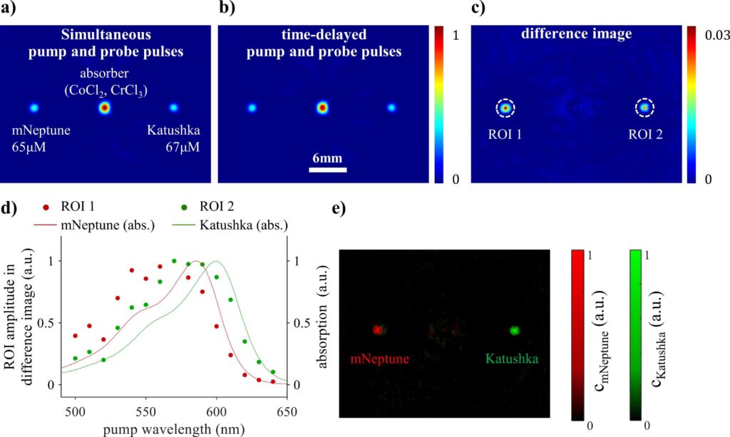
Probing the photophysical properties of fluorescent proteins using photoacoustic pump-probe spectroscopy and imaging

Author(s): F. Ghane Golmohamadi, A. Mehmood, H. T. Phan, F.-J. Schmitt, and J. Laufer


ABSTRACT

Pump-probe excitation of fluorophores has been shown to overcome the limitations of conventional multiwavelength imaging and linear unmixing approaches by providing fluorophore-specific contrast whilst eliminating the dominant background signal of endogenous chromophores. In this study, methods for generating pump-probe signals and images are investigated that rely on changing 1) the pump wavelength whilst keeping the probe wavelength fixed, 2) the probe wavelength whilst keeping the pump wavelength fixed, and 3) the time delay between the pump and probe pulse. Time-resolved PA signals were generated in purified solutions of genetically expressed red fluorescent proteins Katushka, mNeptune, and mCardinal in a cuvette. Spectra of the difference signal amplitude were found to correlate with the absorption and emission spectra. The difference signal plotted as a function of time delay also showed characteristic features for each protein. To demonstrate the capability of multiplexed imaging, the spatial distributions of Katushka and mNeptune were recovered from 2D difference images of a phantom. This study demonstrates that methods based on pump-probe excitation can be used to probe the photophysical properties of fluorophores. By detecting changes in these properties due to a stimulant, such as pH, the methods may find application in biosensing of the cellular microenvironment.

Read the full publication →

Citation

F. Ghane Golmohamadi, A. Mehmood, H. T. Phan, F.-J. Schmitt, and J. Laufer, "Probing the photophysical properties of fluorescent proteins using photoacoustic pump-probe spectroscopy and imaging," Photoacoustics, vol. 44, art. 100738, 2025, doi: 10.1016/j.pacs.2025.100738.