Recent Posts

Single Wavelength Reconstruction of Optical Absorption Coefficient and Elastic Modulus Utilizing Photoacoustic Tomography

Author(s): L. Gong, C. Cai, F. Lin, Z. Xu, Z. Wang, H. Li, and Y. Liu

ABSTRACT

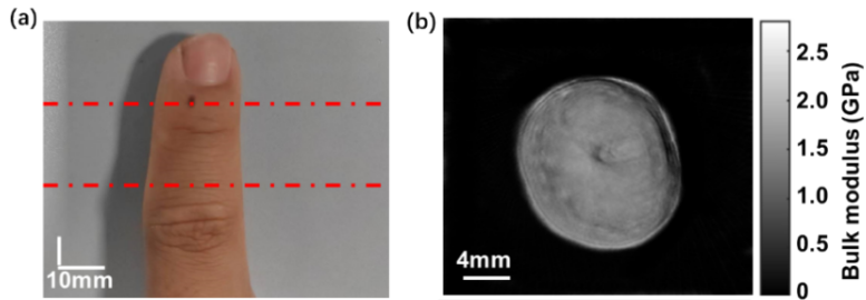
In this study, we utilized a home-made photoacoustic tomography system to recover optical absorption coefficient and elastic modulus under single wavelength. To reconstruct the optical absorption coefficient from photoacoustic measurements, we amalgamate the finite element solutions to the photoacoustic wave equation with MC simulation. Upon determining the absorption coefficient, the elastic modulus reconstruction relies on the analysis of the photoacoustic elastic tomography wave equation. In ex vivo trials, quantitative pencil lead and porcine liver tissue showed an absorption coefficient of 5015±5mm-1 and 0.236±0.01mm-1 at the wavelength of 800nm, respectively, while their elastic modulus parameters were 45.5±0.5GPa and 2.5±0.2GPa. Additionally, we implemented this methodology on in vivo three-dimensional quantitative finger imaging, obtaining the distribution of three-dimensional vascular absorption coefficients and elasticity modulus of the finger at the wavelength of 800nm. From these results, we confirm the method’s capability to discern differences in optical and acoustic properties between normal and abnormal tissues.

Read the full publication →

Citation

L. Gong, C. Cai, F. Lin, Z. Xu, Z. Wang, H. Li, and Y. Liu, Single Wavelength Reconstruction of Optical Absorption Coefficient and Elastic Modulus Utilizing Photoacoustic Tomography (SSRN preprint). 2024. doi: 10.2139/ssrn.4852482.
3D Frequency-Domain Full Waveform Inversion for Whole-Breast Imaging With a Multi-Row Ring Array

Author(s): R. Ali, G. Jin, M. Singh, T. Mitcham, and N. Duric


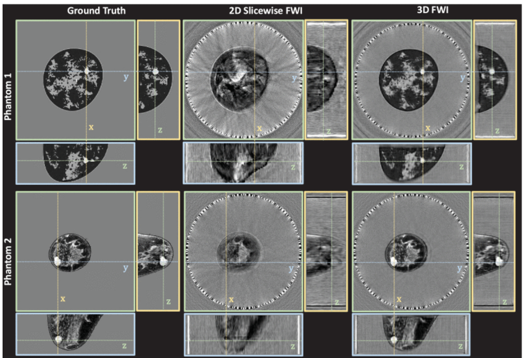
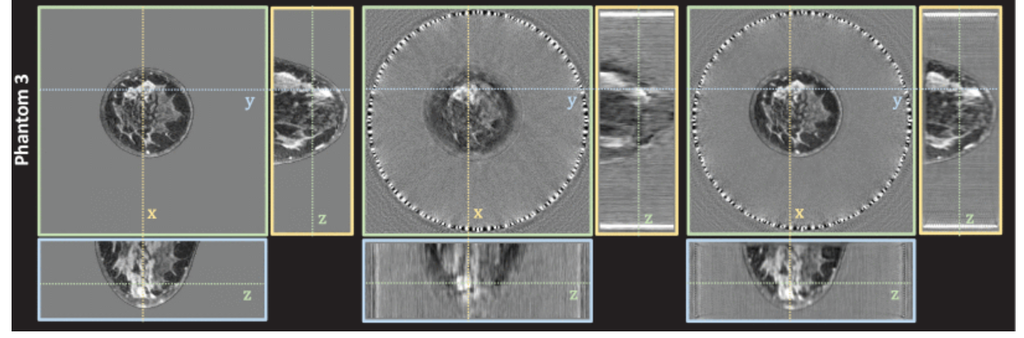
Comparison of 2D slicewise to 3D FWI with simulated multi-row ring array. Cylindrical wave transmits from a multi-row ring-array (32 rows; 256 elements per row; 22 cm diameter; 2.4 mm between rows) were simulated in three different numerical breast phantoms. Orthographic slice views of the reconstructed volumes intersect at suspicious high sound speed masses in each phantom (cancers in phantoms 1 and 2, and dense breast tissue in phantom 3). Each sound speed images is displayed in grayscale from 1400 to 1600 m/s: (Left Column) ground-truth sound speed image; (Middle Column) volume reconstructed using 2D slicewise FWI; (Right Column) volume reconstructed using 3D FWI.

ABSTRACT

For ring-array ultrasound tomography, two-dimensional frequency-domain full waveform inversion is the clinical gold standard for high-resolution imaging of the breast. While yielding high-resolution images in the plane of the ring-array, the resulting slice-wise approach yields lower resolution out of plane when used to reconstruct the full volume. Instead, this work proposes a fully three-dimensional full-waveform inversion based on a multi-row ring-array transducer to improve out-of-plane resolution, while using cylindrical-wave transmissions to minimize acquisition and reconstruction time. For each numerical breast phantom tested, the root-mean-square error of three-dimensional full-waveform inversion is less than that of two-dimensional slice-wise full-waveform inversion by 6.3-13.7 m/s.

Read the full publication →

Citation

R. Ali, G. Jin, M. Singh, T. Mitcham, and N. Duric, “3D Frequency-Domain Full Waveform Inversion for Whole-Breast Imaging With a Multi-Row Ring Array,” IEEE Open Journal of Ultrasonics, Ferroelectrics, and Frequency Control, vol. 5, pp. 77-81, 2025, doi: 10.1109/OJUFFC.2025.3570253.

Enhanced clinical photoacoustic vascular imaging through a skin localization network and adaptive weighting

Author(s): Chuqin Huang, Emily Zheng, Wenhan Zheng, Huijuan Zhang, Yanda Cheng, Xiaoyu Zhang, Varun Shijo, Robert W. Bing, Isabel Komornicki, Linda M. Harris, Ermelinda Bonaccio, Kazuaki Takabe, Emma Zhang, Wenyao Xu, Jun Xia


ABSTRACT

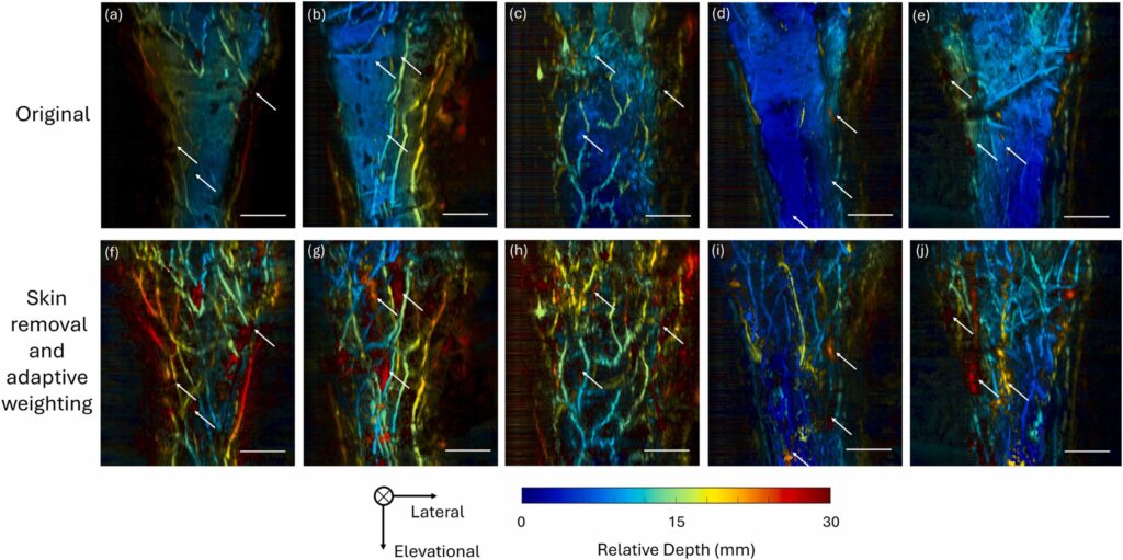
Photoacoustic tomography (PAT) is an emerging imaging modality with widespread applications in both preclinical and clinical studies. Despite its promising capabilities to provide high-resolution images, the visualization of vessels might be hampered by skin signals and attenuation in tissues. In this study, we have introduced a framework to retrieve deep vessels. It combines a deep learning network to segment skin layers and an adaptive weighting algorithm to compensate for attenuation. Evaluation of enhancement using vessel occupancy metrics and signal-to-noise ratio (SNR) demonstrates that the proposed method significantly recovers deep vessels across various body positions and skin tones. These findings indicate the method’s potential to enhance quantitative analysis in preclinical and clinical photoacoustic research.

Read the full publication →

Citation

C. Huang, E. Zheng, W. Zheng, H. Zhang, Y. Cheng, X. Zhang, V. Shijo, R. W. Bing, I. Komornicki, L. M. Harris, E. Bonaccio, K. Takabe, E. Zhang, W. Xu, and J. Xia, "Enhanced clinical photoacoustic vascular imaging through a skin localization network and adaptive weighting," Photoacoustics, vol. 42, art. 100690, 2025, doi: 10.1016/j.pacs.2025.100690.

Utility of Low-Cost Multichannel Data Acquisition System for Photoacoustic Computed Tomography

Author(s): Zafar, Mohsin, Rayyan Manwar, Seyed Mohsen Ranjbaran, and Kamran Avanaki


ABSTRACT

Typically, multi-single-element photoacoustic computed tomography (PACT) systems utilize numerous ultrasound transducers arranged in cylindrical or hemispherical configurations for detection, combined with a single diffuse light source or multiple sparse light sources to illuminate the imaging target. While these systems produce high-quality 3D PA images, they require complex, multi-channel data acquisition (DAQ) systems to acquire data from all transducers. These DAQ systems are often bulky and expensive, significantly limiting the clinical translation of PACT systems for patient care. In this study, we evaluated the feasibility of using a compact and cost-effective Texas Instruments analog front-end DAQ module for multi-single-element PACT systems. By imaging a simple 3D phantom, we demonstrated the capability of this affordable DAQ board, with reconstructed images showing promise for practical and economical solutions in PACT systems. This advancement paves the way for broader applications of PACT in both research and clinical settings.

Read the full publication →

Citation

M. Zafar, R. Manwar, S. M. Ranjbaran, and K. Avanaki, "Utility of Low-Cost Multichannel Data Acquisition System for Photoacoustic Computed Tomography," Photonics, vol. 12, no. 4, art. 385, 2025, doi: 10.3390/photonics12040385.

Signal-domain speed-of-sound correction for ring-array-based photoacoustic tomography

Author(s): Daohuai Jiang, Hengrong Lan, Shangqing Tong, Xianzeng Zhang, Fei Gao


ABSTRACT

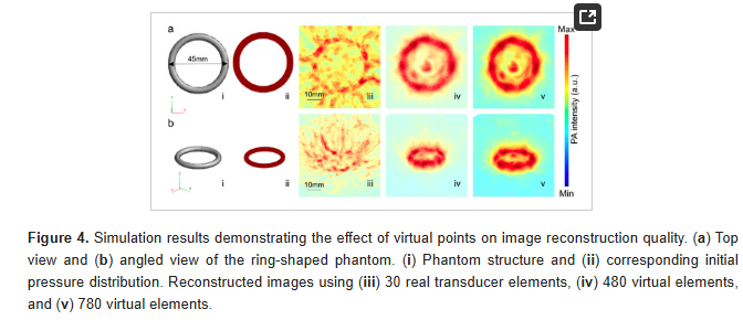
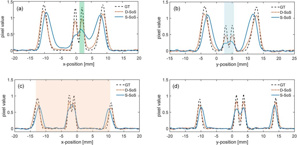
Photoacoustic imaging combines the advantages of optical and acoustic imaging, making it a promising tool in biomedical imaging. Photoacoustic tomography (PAT) reconstructs images by solving the inverse problem from detected photoacoustic waves to initial pressure map. The heterogeneous speed of sound (SoS) distribution in biological tissue significantly affects image quality, as uncertain SoS variations can cause image distortions. Previously reported dual-speed-of-sound (dual-SoS) imaging methods effectively address these distortions by accounting for the SoS differences between tissues and the coupling medium. However, these methods require recalculating the distribution parameters of the SoS for each frame during dynamic imaging, which is highly time-consuming and poses a significant challenge for achieving real-time dynamic dual-SoS PAT imaging. To address this issue, we propose a signal-domain dual-SoS correction method for PAT image reconstruction. In this method, two distinct SoS regions are differentiated by recognizing the photoacoustic signal features of the imaging target's contours. The signals are then corrected based on the respective SoS values, enabling signal-domain-based dual-SoS dynamic real-time PAT imaging. The proposed method was validated through numerical simulations and in-vivo experiments of human finger. The results show that, compared to the single-SoS reconstruction method, the proposed approach produces higher-quality images, achieving the resolution error by near 12 times and a 30 % increase in contrast. Furthermore, the method enables dual-SoS dynamic real-time PAT reconstruction at 10 fps, which is 187.22 % faster than existing dual-SoS reconstruction methods, highlighting its feasibility for dynamic PAT imaging of heterogeneous tissues.

Read the full publication →

Citation

D. Jiang, H. Lan, S. Tong, X. Zhang, and F. Gao, "Signal-domain speed-of-sound correction for ring-array-based photoacoustic tomography," Photoacoustics, vol. 44, art. 100735, 2025, doi: 10.1016/j.pacs.2025.100735.

Advancing precision photothermal therapy byintegrating armored gold nanostars with real-timephotoacoustic thermometry and imaging

Author(s): Aidan J. Canning, Tri Vu1, Luca Menozzi, Paul Klippel, Xinrong Chen, Jianing Chen,Theresa Charity, Khang Hoang, Joseph J. Yang, Yun Jing, Gregory M. Palmer5,Junjie Yao1, Tuan Vo-Dinh


ABSTRACT

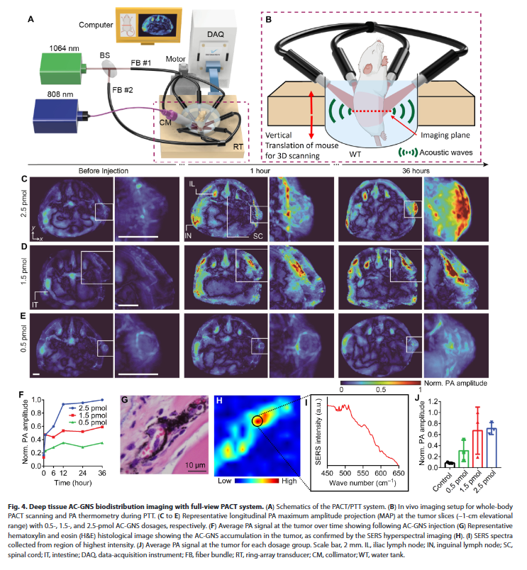
Nanoparticle-mediated photothermal therapy (PTT) is a promising strategy for cancer treatment; however,nanoparticle instability and lack of precise imaging tools for real-time temperature monitoring during therapyand nanoparticle tracking have hindered investigations in animal models. To address these critical issues, we pres-ent a theranostic platform that seamlessly integrates armored core–gold nanostar (AC-GNS)–mediated PTT withfull-view photoacoustic computed tomography (PACT), enabling nanoparticle tracking and real-time imaging-guided PTT in deep tissues. The AC-GNS platform delivered exceptional photostability and thermal resilience be-yond those of conventional nanoparticles while serving as a high-performance contrast agent for PACT and aphotothermal transducer for PTT. Integrating AC-GNS–mediated PTT with noninvasive PACT enabled whole-bodynanoparticle tracking, PTT treatment monitoring via thermal imaging, and thermal dose determination, culminat-ing in a 100% survival rate in a murine bladder cancer model without long-term treatment-related toxicity. Thistheranostic platform lays the foundation for broader research applications and provides opportunities for ad-vancing solid tumor treatment and response assessment research.

Read the full publication →

Citation

A. J. Canning, T. Vu, L. Menozzi, P. Klippel, X. Chen, J. Chen, T. Charity, K. Hoang, J. J. Yang, Y. Jing, G. M. Palmer, and T. Vo-Dinh, "Advancing precision photothermal therapy by integrating armored gold nanostars with real-time photoacoustic thermometry and imaging," Science Advances, vol. 11, no. 33, art. eadx6350, 2025, doi: 10.1126/sciadv.adx6350.

Investigating the Impact of Chronical Prenatal Alcohol Exposure on Fetal Vascular Development Across Pregnancy Stages Using Photoacoustic Tomography

Author(s): Hao Yang, Md Farhan Tanvir, Huabei Jiang


ABSTRACT

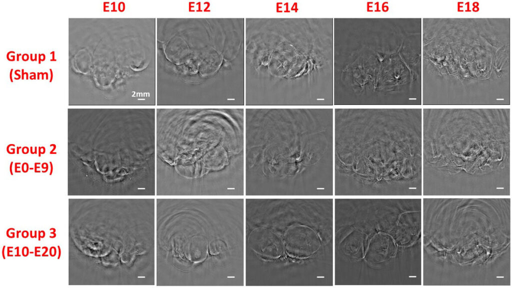
This study investigates the effects of chronic prenatal alcohol exposure on fetal brain vascular development in mice using photoacoustic tomography. Key vascular parameters such as vessel diameter, density, and oxygen saturation were analyzed from embryonic Days 10 to 20. Results demonstrate significant vascular disruptions in alcohol-exposed groups compared to controls, highlighting the vulnerability of fetal development to alcohol exposure and the utility of photoacoustic tomography in detecting these changes.

Read the full publication →

Citation

H. Yang, M. F. Tanvir, and H. Jiang, "Investigating the Impact of Chronical Prenatal Alcohol Exposure on Fetal Vascular Development Across Pregnancy Stages Using Photoacoustic Tomography," Journal of Biophotonics, vol. 18, no. 2, art. e202400410, 2025, doi: 10.1002/jbio.202400410.

A comparative study of plasmonic nanoparticles for targeted photothermal therapy of melanoma tumors using various irradiation modes

Author(s): L. Mikhailova, E. Vysotina, M. Timofeeva, E. Kopoleva, V. Gulinyan, O. Pashina, K. Arabuli, O. Gusliakova, E. Prikhozhdenko, X. Qi, A. Petrov, E. Ageev, M. Petrov, C. De Angelis, M. Durymanov, G. Sukhorukov, and M. V. Zyuzin


ABSTRACT

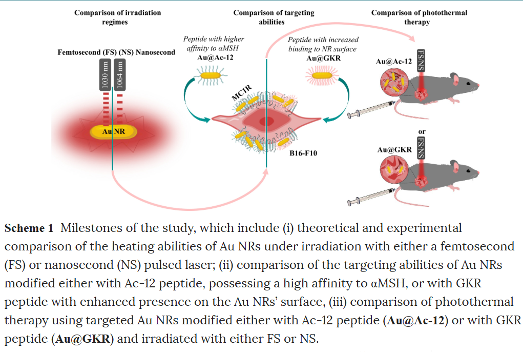
Melanoma, a highly malignant and complex form of cancer, has increased in global incidence, with a growing number of new cases annually. Active targeting strategies, such as leveraging the α-melanocyte-stimulating hormone (αMSH) and its interaction with the melanocortin 1 receptor (MC1R) overexpressed in melanoma cells, enhance the concentration of therapeutic agents at tumor sites. For instance, targeted delivery of plasmonic light-sensitive agents and precise hyperthermia management provide an effective, minimally invasive treatment for tumors. In this work, we present a comparative study on targeted photothermal therapy (PTT) using plasmonic gold nanorods (Au NRs) as a robust and safe nanotool to reveal how key treatment parameters affect therapy outcomes. Using an animal model (B16-F10) of melanoma tumors, we compare the targeting abilities of Au NRs modified with two different MC1R agonists, either closely mimicking the αMSH sequence or providing a superior functionalization extent of Au NRs (4.5% (w/w) versus 1.8% (w/w)), revealing 1.6 times better intratumoral localization. Following theoretical and experimental assessments of the heating capabilities of the developed Au NRs under laser irradiation in either the femtosecond (FS)- or nanosecond (NS)- pulsed regime, we perform targeted PTT employing two types of peptide-modified Au NRs and compare therapeutic outcomes revealing the most appropriate PTT conditions. Our investigation reveals greater heat release from Au NRs under irradiation with FS laser, due to the relaxation rates of the electron and phonon temperatures dissipating in the surrounding, which correlates with a more pronounced 17.6 times inhibition of tumor growth when using FS-pulsed regime.

Read the full publication →

Citation

L. Mikhailova, E. Vysotina, M. Timofeeva, E. Kopoleva, V. Gulinyan, O. Pashina, K. Arabuli, O. Gusliakova, E. Prikhozhdenko, X. Qi, A. Petrov, E. Ageev, M. Petrov, C. De Angelis, M. Durymanov, G. Sukhorukov, and M. V. Zyuzin, "A comparative study of plasmonic nanoparticles for targeted photothermal therapy of melanoma tumors using various irradiation modes," Light: Advanced Manufacturing, vol. 6, no. 1, art. 005, 2025, doi: 10.37188/lam.2025.005.

Rotational ultrasound and photoacoustic tomography of the human body

Author(s): Yang Zhang, Shuai Na, Jonathan J. Russin, Karteekeya Sastry, Li Lin, Junfu Zheng, Yilin Luo, Xin Tong, Yujin An, Peng Hu, Konstantin Maslov, Tze-Woei Tan, Charles Y. Liu, Lihong V. Wang


ABSTRACT

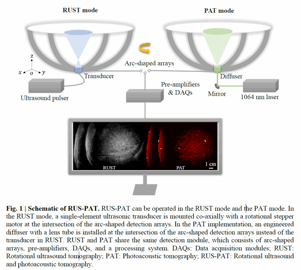
Imaging the human body's morphological and angiographic information is essential for diagnosing, monitoring, and treating medical conditions. Ultrasonography performs the morphological assessment of the soft tissue based on acoustic impedance variations, whereas photoacoustic tomography (PAT) can visualize blood vessels based on intrinsic hemoglobin absorption. Three-dimensional (3D) panoramic imaging of the vasculature is generally not practical in conventional ultrasonography with limited field-of-view (FOV) probes, and PAT does not provide sufficient scattering-based soft tissue morphological contrast. Complementing each other, fast panoramic rotational ultrasound tomography (RUST) and PAT are integrated for hybrid rotational ultrasound and photoacoustic tomography (RUS-PAT), which obtains 3D ultrasound structural and PAT angiographic images of the human body quasi-simultaneously. The RUST functionality is achieved in a cost-effective manner using a single-element ultrasonic transducer for ultrasound transmission and rotating arc-shaped arrays for 3D panoramic detection. RUST is superior to conventional ultrasonography, which either has a limited FOV with a linear array or is high-cost with a hemispherical array that requires both transmission and receiving. By switching the acoustic source to a light source, the system is conveniently converted to PAT mode to acquire angiographic images in the same region. Using RUS-PAT, we have successfully imaged the human head, breast, hand, and foot with a 10 cm diameter FOV, submillimeter isotropic resolution, and 10 s imaging time for each modality. The 3D RUS-PAT is a powerful tool for high-speed, 3D, dual-contrast imaging of the human body with potential for rapid clinical translation.

Read the full publication →

Citation

Y. Zhang, S. Na, J. J. Russin, K. Sastry, L. Lin, J. Zheng, Y. Luo, X. Tong, Y. An, P. Hu, K. Maslov, T.-W. Tan, C. Y. Liu, and L. V. Wang, "Rotational ultrasound and photoacoustic tomography of the human body," Nature Biomedical Engineering, 2026, doi: 10.1038/s41551-025-01603-5.

3D protoacoustic radiography: A proof of principle study

Author(s): Prabodh K Pandey, Gilberto Gonzalez, Kristina Bjegovic, Leshan Sun, Yong Chen, Liangzhong Xiang


ABSTRACT

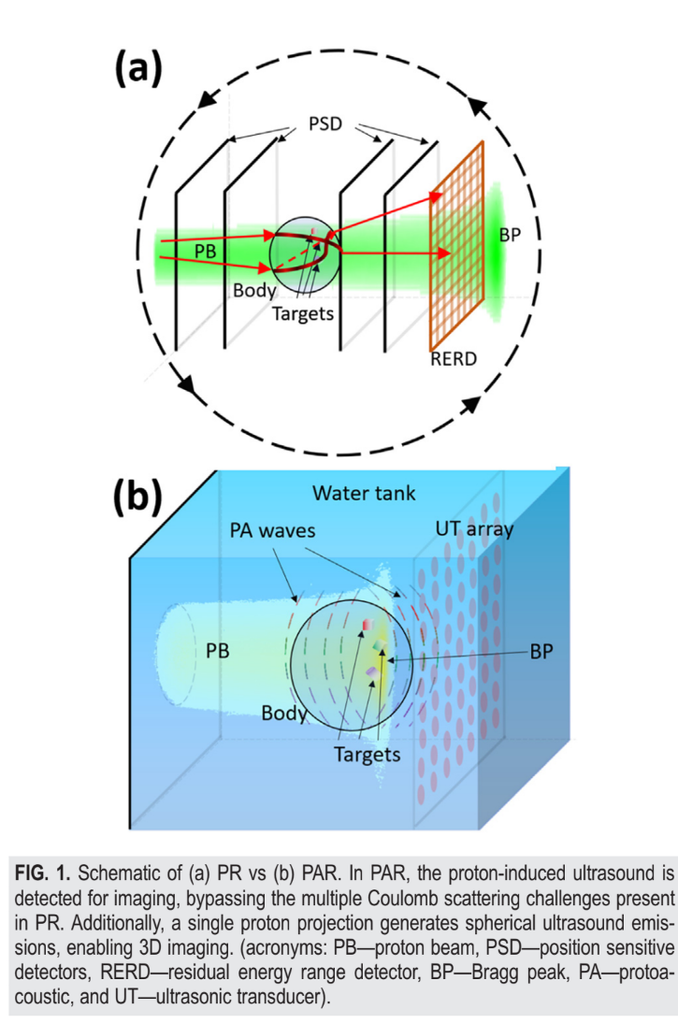
We propose protoacoustic radiography (PAR), an imaging modality combining proton excitation and acoustic detection for three-dimensional (3D) imaging from a single proton projection. PAR avoids the effect of multiple Coulomb scattering in imaging by detecting ultrasound. Proton-induced acoustic waves propagate spherically, enabling 3D imaging from a single projection. Additionally, the distinctive feature of proton beams—concentrating energy deposition primarily at the Bragg peak—allows for precise depth-selectivity through proton energy tuning. We performed PAR using clinical proton machines, and our results demonstrate the capability of PAR to reconstruct targets at various depths (between ∼20 and 23 cm) with an axial resolution of 1.3 mm by fully leveraging the Bragg peak and by tuning the kinetic energy of the proton beam. PAR offers opportunities for precise structural determination with protons both in biomedicine and nondestructive testing.

Read the full publication →

Citation

P. K. Pandey, G. Gonzalez, K. Bjegovic, L. Sun, Y. Chen, and L. Xiang, “3D protoacoustic radiography: A proof of principle study,” Applied Physics Letters, vol. 126, no. 7, art. 074102, 2025, doi: 10.1063/5.0239878.