
Author(s): R. Ali, G. Jin, M. Singh, T. Mitcham, and N. Duric
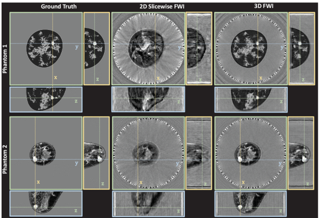
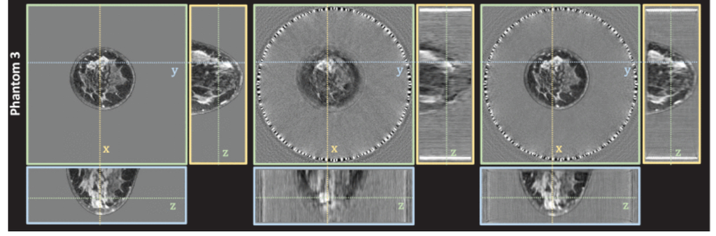
Comparison of 2D slicewise to 3D FWI with simulated multi-row ring array. Cylindrical wave transmits from a multi-row ring-array (32 rows; 256 elements per row; 22 cm diameter; 2.4 mm between rows) were simulated in three different numerical breast phantoms. Orthographic slice views of the reconstructed volumes intersect at suspicious high sound speed masses in each phantom (cancers in phantoms 1 and 2, and dense breast tissue in phantom 3). Each sound speed images is displayed in grayscale from 1400 to 1600 m/s: (Left Column) ground-truth sound speed image; (Middle Column) volume reconstructed using 2D slicewise FWI; (Right Column) volume reconstructed using 3D FWI.
ABSTRACT
For ring-array ultrasound tomography, two-dimensional frequency-domain full waveform inversion is the clinical gold standard for high-resolution imaging of the breast. While yielding high-resolution images in the plane of the ring-array, the resulting slice-wise approach yields lower resolution out of plane when used to reconstruct the full volume. Instead, this work proposes a fully three-dimensional full-waveform inversion based on a multi-row ring-array transducer to improve out-of-plane resolution, while using cylindrical-wave transmissions to minimize acquisition and reconstruction time. For each numerical breast phantom tested, the root-mean-square error of three-dimensional full-waveform inversion is less than that of two-dimensional slice-wise full-waveform inversion by 6.3-13.7 m/s.
Citation
R. Ali, G. Jin, M. Singh, T. Mitcham, and N. Duric, “3D Frequency-Domain Full Waveform Inversion for Whole-Breast Imaging With a Multi-Row Ring Array,” IEEE Open Journal of Ultrasonics, Ferroelectrics, and Frequency Control, vol. 5, pp. 77-81, 2025, doi: 10.1109/OJUFFC.2025.3570253.