
Author(s): Li Lin, Xin Tong, Susana Cavallero, Yide Zhang, Shuai Na, Rui Cao, Tzung K. Hsiai, and Lihong V. Wang
ABSTRACT
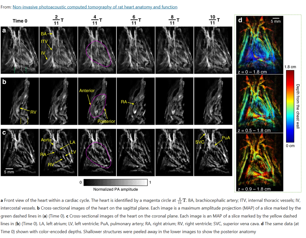
Photoacoustic computed tomography (PACT) combines optical contrast with acoustic-resolution imaging, but non-invasive cardiac imaging with this technique has been limited by signal loss and distortion caused by the chest wall. This work presents a 3D-PACT platform designed to overcome that limitation, using an optimized illumination and detection scheme to image rat hearts non-invasively — without surgical exposure of the chest — capturing a full cardiac scan in about 10 seconds. The system reveals both cardiac anatomy and intracardiac blood flow, gated to the animal’s ECG signal to freeze motion at each point in the cardiac cycle. Applied across healthy, hypertensive, and obese rat models, the technique distinguished differences in chamber size, wall thickness, and hemodynamics between groups, pointing toward potential future use in non-invasive, high-speed cardiac imaging of human neonates.
Citation
L. Lin, X. Tong, S. Cavallero, Y. Zhang, S. Na, R. Cao, T. K. Hsiai, and L. V. Wang, “Non-Invasive Photoacoustic Computed Tomography of Rat Heart Anatomy and Function,” Light: Science & Applications, vol. 12, 2023, doi: 10.1038/s41377-022-01053-7.