Recent Posts

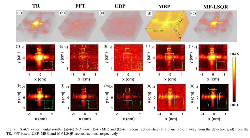
Model-Based 3-D X-Ray Induced Acoustic Computerized Tomography

Authors: Prabodh Kumar Pandey; Siqi Wang; Leshan Sun; Lei Xing; Liangzhong Xiang

ABSTRACT

X-ray-induced acoustic (XA) computerized tomography (XACT) is an evolving imaging technique that aims to reconstruct the X-ray energy deposition from XA measurements. Main challenges in XACT are the poor signal-to-noise ratio and limited field-of-view, which cause artifacts in the images. We demonstrate the efficacy of model-based (MB) algorithms for 3-D XACT and compare with the traditional algorithms. The MB algorithms are based on the matrix free approach for regularized-least-squares minimization corresponding to XACT. The matrix-free-LSQR (MF-LSQR) and the noniterative model-backprojection (MBP) reconstructions were evaluated and compared with universal backprojection (UBP), time-reversal (TR), and fast-Fourier transform (FFT)-based reconstructions for numerical and experimental XACT datasets. The results demonstrate the capability of the MF-LSQR algorithm to reduce noisy artifacts thus yielding better reconstructions. MBP and MF-LSQR algorithms perform particularly well with the experimental XACT dataset, where noise in signals significantly affects the reconstruction of the target in UBP and FFT-based reconstructions. The TR reconstruction for experimental XACT is comparable to MF-LSQR, but takes thrice as much time and filters the frequency components greater than maximum frequency supported by the grid, resulting loss of resolution. The MB algorithms are able to overcome the challenges in XACT and hence are vital for the clinical translation of XACT.

Read the full publication →

Citation

P. K. Pandey, S. Wang, L. Sun, L. Xing, and L. Xiang, “Model-Based 3-D X-Ray Induced Acoustic Computerized Tomography,” IEEE Transactions on Radiation and Plasma Medical Sciences, vol. 7, no. 5, p. 532, 2023, doi: 10.1109/TRPMS.2023.3238017.
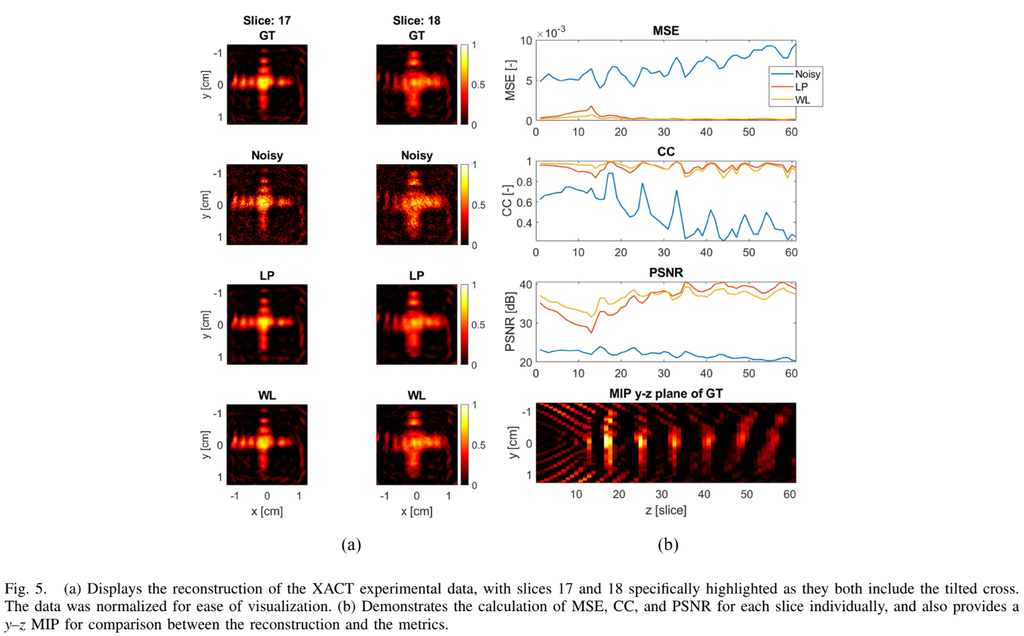
Discrete Wavelet Transformation for the Sensitive Detection of Ultrashort Radiation Pulse with Radiation-Induced Acoustics

Authors: Rick Van Bergen, Leshan Sun, Prabodh Kumar Pandey, Siqi Wang, Kristina Bjegovic, Gilberto Gonzalez, Yong Chen, Richard Lopata, Liangzhong Xiang


ABSTRACT

Radiation-induced acoustics (RIA) shows promise in advancing radiological imaging and radiotherapy dosimetry methods. However, RIA signals often require extensive averaging to achieve reasonable signal-to-noise ratios, which increases patient radiation exposure and limits real-time applications. Therefore, this article proposes a discrete wavelet transform (DWT)-based filtering approach to denoise the RIA signals and avoid extensive averaging. The algorithm was benchmarked against low-pass filters and tested on various types of RIA sources, including low-energy X-rays, high-energy X-rays, and protons. The proposed method significantly reduced the required averages (1000 times less averaging for low-energy X-ray RIA, 32 times less averaging for high-energy X-ray RIA, and four times less averaging for proton RIA) and demonstrated robustness in filtering signals from different sources of radiation. The coif5 wavelet in conjunction with the sqtwolog threshold selection algorithm yielded the best results. The proposed DWT filtering method enables high-quality, automated, and robust filtering of RIA signals, with a performance similar to low-pass filtering, aiding in the clinical translation of radiation-based acoustic imaging for radiology and radiation oncology.

Read the full publication →

Citation

R. Van Bergen, L. Sun, P. K. Pandey, S. Wang, K. Bjegovic, G. Gonzalez, Y. Chen, R. Lopata, and L. Xiang, “Discrete Wavelet Transformation for the Sensitive Detection of Ultrashort Radiation Pulse With Radiation-Induced Acoustics,” IEEE Transactions on Radiation and Plasma Medical Sciences, vol. 8, no. 1, p. 76, 2024, doi: 10.1109/TRPMS.2023.3314339.

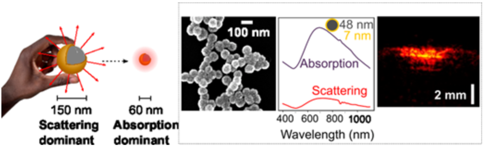
MONODISPERSE SUB-100 NM AU NANOSHELLS FOR LOW-FLUENCE DEEP-TISSUE PHOTOACOUSTIC IMAGING

Author(s): Luis D. B. Manuel, Vinion Devpaul Vincely, Carolyn Bayer, and Kevin M. McPeak


ABSTRACT

Nanoparticles with high absorption cross sections will advance therapeutic and bioimaging nanomedicine technologies. While Au nanoshells have shown great promise in nanomedicine, state-of-the-art synthesis methods result in scattering-dominant particles, mitigating their efficacy in absorption-based techniques that leverage the photothermal effect, such as photoacoustic (PA) imaging. We introduce a highly reproducible synthesis route to monodisperse sub-100 nm Au nanoshells with an absorption-dominant optical response. Au nanoshells with 48 nm SiO2 cores and 7 nm Au shells show a 14-fold increase in their volumetric absorption coefficient compared to commercial Au nanoshells with dimensions commonly used in nanomedicine. PA imaging with Au nanoshell contrast agents showed a 50% improvement in imaging depth for sub-100 nm Au nanoshells compared with the smallest commercially available nanoshells in a turbid phantom. Furthermore, the high PA signal at low fluences, enabled by sub-100 nm nanoshells, will aid the deployment of low-cost, low-fluence light-emitting diodes for PA imaging.

Read the full publication →

Citation

L. D. B. Manuel, V. D. Vincely, C. L. Bayer, and K. M. McPeak, “Monodisperse Sub-100 nm Au Nanoshells for Low-Fluence Deep-Tissue Photoacoustic Imaging,” ACS Nano Letters, 2023, doi: 10.1021/acs.nanolett.3c01696.

SIZE-TUNABLE ICG-BASED CONTRAST AGENT PLATFORM FOR TARGETED NEAR-INFRARED PHOTOACOUSTIC IMAGING

Author(s): Shrishti Singh, Giovanni Giammanco, Chih-Hsiang Hu, Joshua Bush, Leandro Soto Cordova, Dylan J Lawrence, Jeffrey L Moran, Parag V Chitnis, Remi Veneziano


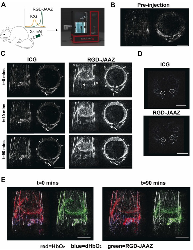
Fig. 6. In vivo imaging using RGD-JAAZ particles. (A) Schematic and photograph of the 3D PAI acquisition on the TriTom™ imaging system. (B) Coronal and axial slabs of the baseline PA signal acquired at 895 nm. (C) Representative coronal and axial slabs of the ICG (left) and RGD-JAAZ particle (right) injections acquired at the peak excitation wavelength (i.e., 800 and 895 nm, respectively) over time. The displayed dynamic range for all images was set based on the baseline scan acquired at the same wavelength. (Legend): a. superficial thoracic vessels, b. liver c. spleen d. intestines e. thoracic vertebrae f. CuSO4 fiducial tube. (D) Axial slabs encompassing the iliac arteries (white ROI) used to calculate the achievable imaging depth for ICG and RGD-JAAZ particles 90-minutes post-injection. (E) Coronal slabs of oxyhemoglobin (red), deoxyhemoglobin (blue), and RGD-JAAZ particles (green) spectral unmixing overlaid on the 800 nm excitation scan of anatomy (white) at 0- and 90-minutes post-injection. Scale bars for all images are 10 mm. (n = 2 female nu/nu nude mice).

ABSTRACT

Near-infrared photoacoustic imaging (NIR-PAI) combines the advantages of optical and ultrasound imaging to provide anatomical and functional information of tissues with high resolution. Although NIR-PAI is promising, its widespread use is hindered by the limited availability of NIR contrast agents. J-aggregates (JA) made of indocyanine green dye (ICG) represents an attractive class of biocompatible contrast agents for PAI. Here, we present a facile synthesis method that combines ICG and ICG-azide dyes for producing contrast agents with tunable size down to 230 nm and direct functionalization with targeting moieties. The ICG-JA platform has a detectable PA signal in vitro that is two times stronger than whole blood and high photostability. The targeting ability of ICG-JA was measured in vitro using HeLa cells. The ICG-JA platform was then injected into mice and in vivo NIR-PAI showed enhanced visualization of liver and spleen for 90 min post-injection with a contrast-to-noise ratio of 2.42.

Read the full publication →

Citation

S. Singh, G. Giammanco, C.-H. Hu, K. Bush, L. Soto Cordova, D. J. Lawrence, J. L. Moran, P. V. Chitnis, and R. Veneziano, “Size-tunable ICG-based contrast agent platform for targeted near-infrared photoacoustic imaging,” Photoacoustics, vol. 29, p. 100437, 2023/02/01/ 2023, doi: https://doi.org/10.1016/j.pacs.2022.100437.

FUNCTIONAL PHOTOACOUSTIC IMAGING FOR PLACENTAL MONITORING: A MINI REVIEW

Author(s): Vinoin Devpaul Vincely, Carolyn L Bayer


ABSTRACT

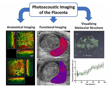
The placenta, a highly vascularized interface between the mother and fetus, undergoes dramatic anatomical and functional changes during pregnancy. These changes occur both during healthy development and adverse pathologies of pregnancy, such as preeclampsia. Abnormal placental development can lead to life-long health impacts on both the mother and child. Photoacoustic (PA) imaging, extensively developed for preclinical imaging applications in oncology and cardiovascular disease, uses optical energy to generate acoustic waves through thermoelastic expansion of light-absorbing chromophores within tissue. Recently, photoacoustic imaging has been used to study preclinical placental anatomy and function. If clinical translation of photoacoustic imaging of the placenta is achieved, the impact on maternal-fetal health could be expansive. This perspective highlights the recent progress in photoacoustic imaging for placental monitoring and discusses the progress needed for human clinical translation.

Read the full publication →

Citation

D. Vincely and C. L. Bayer, “Functional photoacoustic imaging for placental monitoring: A mini review,” IEEE Transactions on Ultrasonics, Ferroelectrics, and Frequency Control, pp. 1-1, 2023, doi: 10.1109/tuffc.2023.3263361.

SPHERICAL-VIEW PHOTOACOUSTIC TOMOGRAPHY FOR MONITORING IN VIVO PLACENTAL FUNCTION

Author(s): Kristie Huda , Chengxi Wu , Jaclyn G. Sider, Carolyn L. Bayer


ABSTRACT

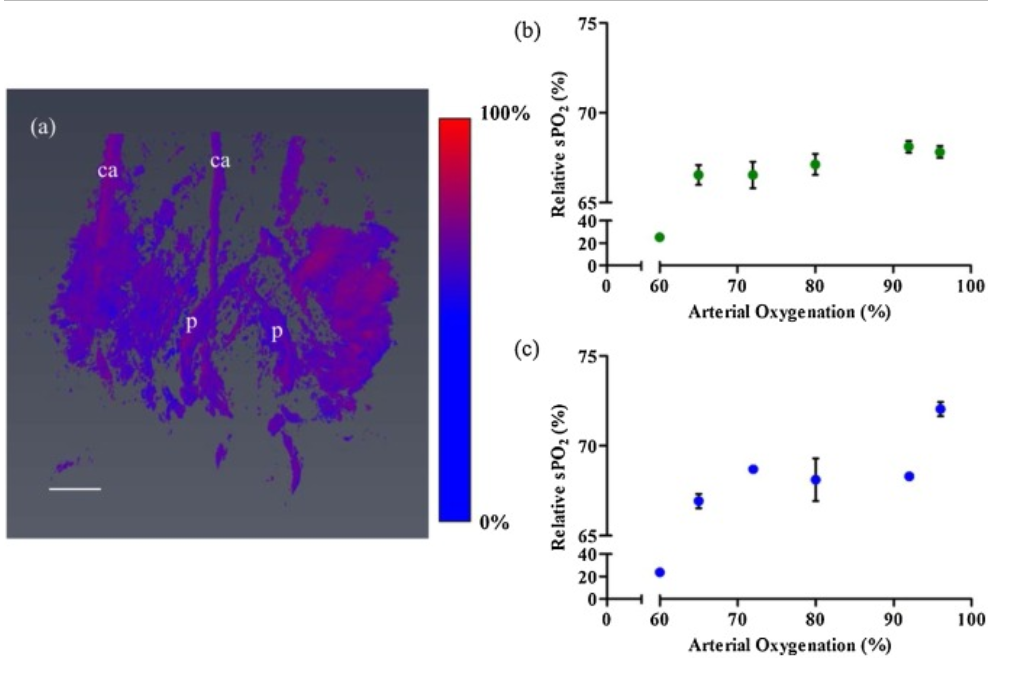
Photoacoustic tomography has great potential to image dynamic functional changes in vivo. Many tomographic systems are built with a circular view geometry, necessitating a linear translation along one axis of the subject to obtain a three-dimensional volume. In this work, we evaluated a prototype spherical view photoacoustic tomographic system which acquires a 3D volume in a single scan, without linear translation. We simultaneously measured relative hemoglobin oxygen saturation in multiple placentas of pregnant mice under oxygen challenge. We also synthesized a folate-conjugated indocyanine green (ICG) contrast agent to image folate kinetics in the placenta. Photoacoustic tomography performed at the wavelength of peak optical absorption of our contrast agent revealed increased ICG signal over time. Through these phantom and in vivo studies, we have demonstrated that the spherical view 3D photoacoustic tomographic system achieves high sensitivity and fast image acquisition, enabling in vivo experiments to assess physiological and molecular dynamics.

Read the full publication →

Citation

K. Huda, C. Wu, J. G. Sider, and C. L. Bayer, "Spherical-view photoacoustic tomography for monitoring in vivo placental function," Photoacoustics, p. 100209, 2020, doi: 10.1016/j.pacs.2020.100209.

NON-INVASIVE 3D PHOTOACOUSTIC TOMOGRAPHY OF ANGIOGRAPHIC ANATOMY AND HEMODYNAMICS OF FATTY LIVERS IN RATS

Author(s): Xin Tong, Li Lin, Peng Hu, Rui Cao, Yang Zhang, Joshua Olick-Gibson,and Lihong V. Wang


ABSTRACT

Non-alcoholic fatty liver disease is the most common liver disorder worldwide, which strongly correlates to obesity, diabetes, and metabolic syndromes. Complementary to mainstream liver diagnostic modalities, photoacoustic tomography (PAT) can provide high-speed images with functional optical contrast. However, PAT has not been demonstrated to study fatty liver anatomy with clear volumetric vasculatures. The livers of multiple rats are non-invasively imaged in vivo using the recently developed 3D PAT platform. The system provides isotropically high spatial resolution in 3D space, presenting clear angiographic structures of rat livers without injecting contrast agents. Furthermore, to quantitatively analyze the difference between the livers of lean and obese rats, the authors measured several PAT features and statistical differences between the two groups are observed. In addition to the anatomy, a time-gated strategy is applied to correct respiration-induced motion artifacts and extracted the hemodynamics of major blood vessels during the breathing cycles. This study demonstrates the capabilities of 3D-PAT to reveal both angiographic anatomy and function in rat livers, providing hematogenous information for fatty liver diagnosis. 3D-PAT, as a new tool for preclinical research, warrants further improvements to be transferred to human pediatric liver imaging.

Read the full publication →

Citation

X. Tong, L. Lin, P. Hu, R. Cao, Y. Zhang, J. Olick-Gibson, and L. V. Wang, “Non-Invasive 3D Photoacoustic Tomography of Angiographic Anatomy and Hemodynamics of Fatty Livers in Rats,” Advanced Science, vol. 10, no. 2, 2023, doi: 10.1002/advs.202205759.

WHOLE-BODY HUMAN ULTRASOUND TOMOGRAPHY

Author(s): D. C. Garrett, J. Xu, D. Oh, Y. Aborahama, G. Ku, K. Maslov, W. W. Tseng, and L. V. Wang


ABSTRACT

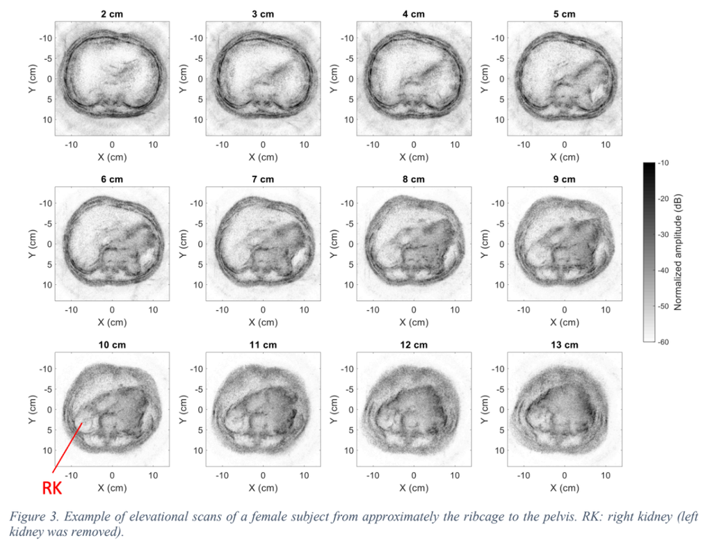
We developed a system for whole-body human ultrasound tomography in reflection andtransmission modes. A custom 512-element ultrasound receiver array with a rotating single-element ultrasound transmitter are used to generate 2D isotropically resolved images across the entire human cross-section. We demonstrate this technique in regions such as the abdomen and legs in healthy volunteers. Compared to handheld-probe-based ultrasonography, this approach provides a substantially larger field of view, depends less on operator training, and obtains quantitative tissue parameter profiles in addition to reflectivity images. Whole-body ultrasound tomography could be valuable in applications such as organ disease screening, image-guided needle biopsy, and treatment monitoring.

Read the full publication →

Citation

D. C. Garrett, J. Xu, D. Oh, Y. Aborahama, G. Ku, K. Maslov, W. W. Tseng, and L. V. Wang, “Whole cross-sectional human ultrasound tomography,” Nature Biomedical Engineering, 2026, doi: 10.1038/s41551-026-01660-4.

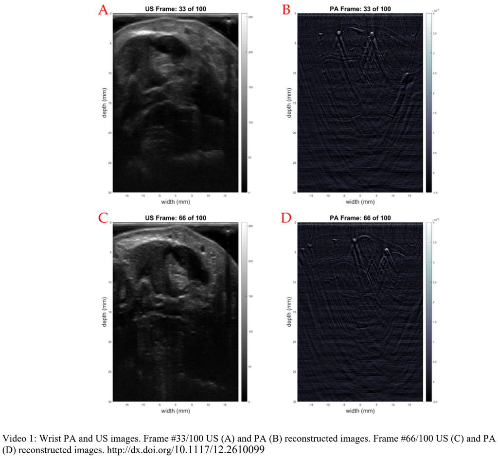
A COMBINED ULTRASOUND AND PHOTOACOUSTIC IMAGING PLATFORM FOR CLINICAL RESEARCH APPLICATIONS

Author(s): Weylan ThompsonHans-Peter BrechtSergey A. ErmilovVassili Ivanov


ABSTRACT

We present the continued development of a clinical ultrasound (US) imaging device with a photoacoustic (PA) mode. The combined USPA imaging platform is designed around a compact US component capable of B-mode, M-mode, color Doppler, and pulsed wave Doppler US imaging modes with a 128-element US probe. The PA mode can support a 256-element PA probe with real-time 2D imaging up to 20 Hz. The PA signals are amplified by a 40 dB pre-amplifier while US mode signals bypass the circuit, resulting in high-quality PA images. We demonstrate the USPA platform’s capabilities using tissue-mimicking phantoms.

Read the full publication →

Citation

W. R. Thompson, D. J. Lawrence, H.-P. Brecht, S. A. Ermilov, and V. Ivanov, A combined ultrasound and photoacoustic imaging platform for clinical research applications (SPIE BiOS). SPIE, 2022. doi: 10.1117/12.2610099.