PhotoSounds OEM Line of products is an ideal starting point for the development of custom systems where the parallel acquisition of multiple channels is required. All our ADCs are streaming and allow the continuous acquisition of data straight to the receiving computer for processing or storage.
PhotoSound’s ADCs are feature-rich, they have multiple electronic and optical trigger inputs as well as programmable outputs that allow the timing control of additional devices. It is possible to combine multiple ADCs in parallel. Simultaneous acquisition of 4096 channels has been realized routinely.
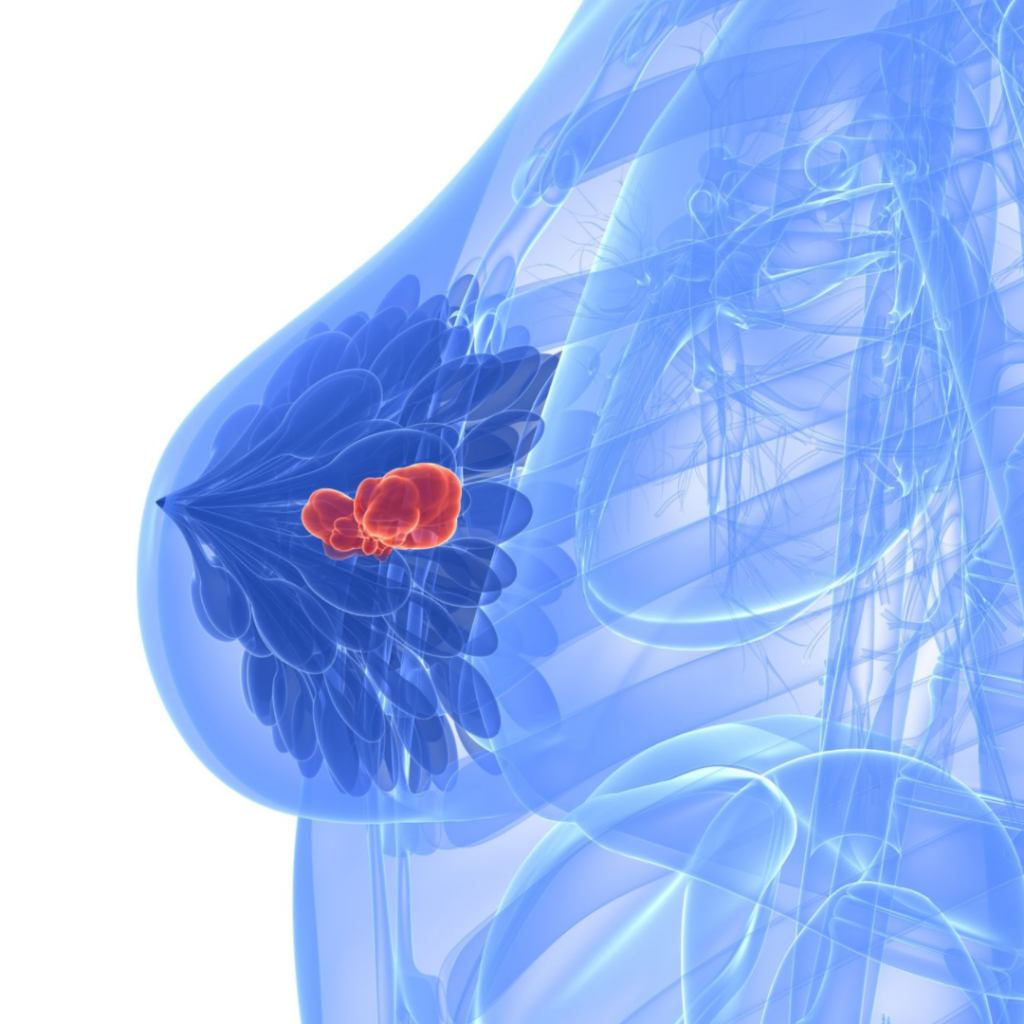
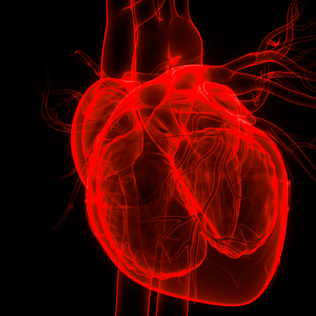
What is Cardiac Imaging?
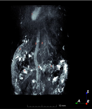
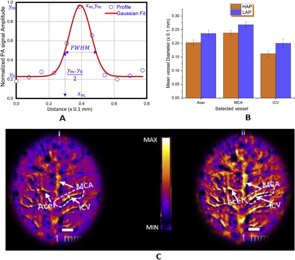
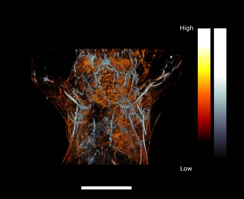
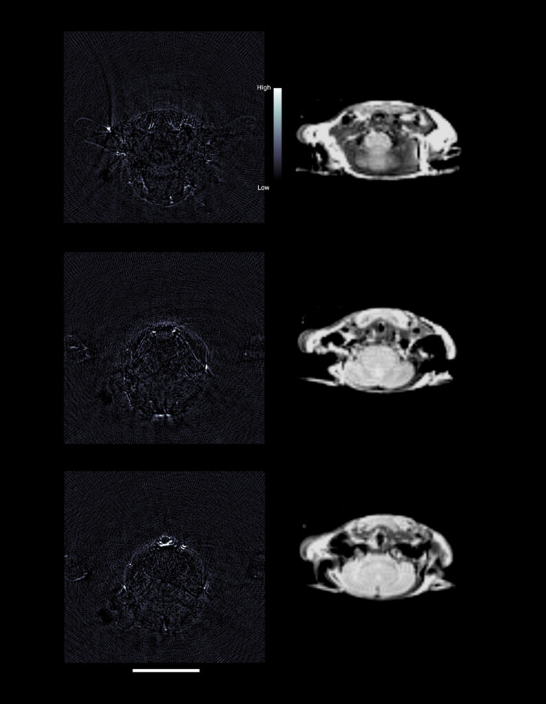
Cardiac Imaging is the use of medical imaging techniques to study the structure and function of the cardiovascular system, focusing on the heart. Photoacoustic tomography has been used to image the heart, blood vessels, and other parts of the cardiovascular system.
PhotoSound Product Used
Non-invasive photoacoustic computed tomography of rat heart anatomy and function