
Author(s): Weylan R. Thompson, Hans-Peter F. Brecht, Vassili Ivanov, Anthony M. Yu, Diego S. Dumani, Dylan J. Lawrence, Stanislav Y. Emelianov, Sergey A. Ermilov
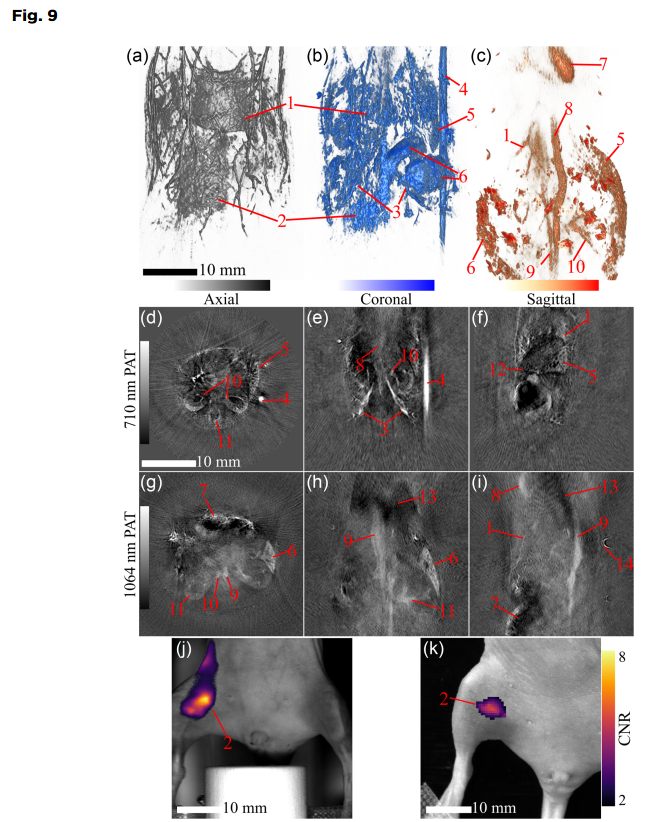
PAT volumes and slices of in vivo mouse scans. The top PAT row shows 3D renders of the PAT volumes scanned at wavelengths (a) 532 nm for superficial structures, (b) 710 nm for deep structures with deoxygenated hemoglobin, and (c) 1064 nm for deep structures with oxygenated hemoglobin in the coronal (ventral) view. The center PAT row has three slices of interest from the 710 nm volume: (d) axial, (e) coronal, and (f) sagittal. The bottom PAT row also has three slices from the 1064-nm volume, with the same view orientations of (g) axial, (h) coronal, and (i) sagittal. The FLI row has FL CNR > 2 maps overlaid on photographs using a TriTom (j) and IVIS Lumina II (k), the color bar is normalized for both instruments. Key anatomical structures and a fiducial are labeled: liver/liver vessels (1), injection site (2), iliac vessels (3), copper sulfate fiducial (4), spleen (5), intestines (6), heart (7), aorta (8), vena cava (9), kidney/kidney vessels (10), vertebra (11), pancreas (12), lungs (13), and hair follicle (14). Scalebars on the 3D (a, b, c), and 2D (d, e, f, g, h, i, j, and k) images are 10 mm. Photoacoustic image color bars are normalized from 0 to 1 from arbitrary units; the FL map color bar represents the CNR of the fluorescent image.
ABSTRACT
To effectively study preclinical animal models, medical imaging technology must be developed with a high enough resolution and sensitivity to perform anatomical, functional, and molecular assessments. Photoacoustic (PA) tomography provides high resolution and specificity, and fluorescence (FL) molecular tomography provides high sensitivity; the combination of these imaging modes will enable a wide range of research applications to be studied in small animals.
We introduce and characterize a dual-modality PA and FL imaging platform using in vivo and phantom experiments.
The imaging platform’s detection limits were characterized through phantom studies that determined the PA spatial resolution, PA sensitivity, optical spatial resolution, and FL sensitivity.
The system characterization yielded a PA spatial resolution of 173 ± 17 μm in the transverse plane and 640 ± 120 μm in the longitudinal axis, a PA sensitivity detection limit not less than that of a sample with absorption coefficient μa = 0.258 cm − 1, an optical spatial resolution of 70 μm in the vertical axis and 112 μm in the horizontal axis, and a FL sensitivity detection limit not <0.9 μM concentration of IR-800. The scanned animals displayed in three-dimensional renders showed high-resolution anatomical detail of organs.
The combined PA and FL imaging system has been characterized and has demonstrated its ability to image mice in vivo, proving its suitability for biomedical imaging research applications.
Citation
W. R. Thompson, H.-P. Brecht, V. Ivanov, A. M. Yu, D. S. Dumani, D. J. Lawrence, S. Y. Emelianov, and S. A. Ermilov, “Characterizing a photoacoustic and fluorescence imaging platform for preclinical murine longitudinal studies,” J. Biomed. Opt., vol. 28, no. 3, p. 036001, 2023. https://doi.org/10.1117/1.JBO.28.3.036001.